2VZX

Structural and spectroscopic characterization of photoconverting fluorescent protein Dendra2


Experimental Data Snapshot

  • Method: X-RAY DIFFRACTION
  • Resolution: 2.00 Å
  • R-Value Free: 0.269 
  • R-Value Work: 0.212 
  • R-Value Observed: 0.215 

wwPDB Validation   3D Report Full Report


This is version 3.1 of the entry. See complete history


Literature

Structural Basis of Enhanced Photoconversion Yield in Green Fluorescent Protein-Like Protein Dendra2.

Adam, V.Nienhaus, K.Bourgeois, D.Nienhaus, G.U.

(2009) Biochemistry 48: 4905

  • DOI: https://doi.org/10.1021/bi900383a
  • Primary Citation of Related Structures:  
    2VZX

  • PubMed Abstract: 

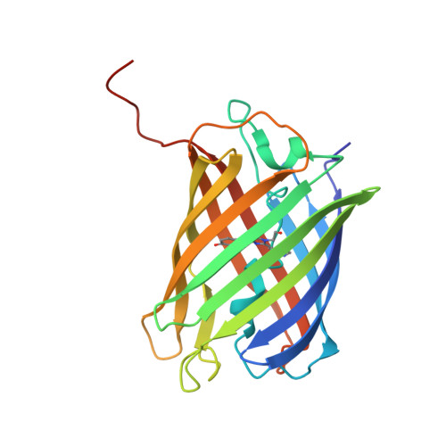
    Dendra2 is an engineered, monomeric GFP-like protein that belongs to a subclass of fluorescent proteins undergoing irreversible photoconversion from a green- to a red-emitting state upon exposure to purple-blue light. This photoinduced process occurs only in the neutral state of the chromophore and is known to result from backbone cleavage accompanied by an extension of the delocalized pi-electron system. We have measured the X-ray structure of the green species of Dendra2 and performed a comprehensive characterization of the optical absorption and fluorescence properties of the protein in both its green and red forms. The structure, which is very similar to those reported for the closely related proteins EosFP and Kaede, revealed a local structural change involving mainly Arg66 and a water molecule W4, which are part of a charged and hydrogen-bonded cluster of amino acids and water molecules next to the chromophore. Unlike in EosFP and Kaede, Arg66 of Dendra2 does not contribute to negative charge stabilization on the imidazolinone ring by hydrogen bonding to the imidazolinone carbonyl. This structural change may explain the blue shift of the absorption and emission bands, as well as the markedly higher pKs of the hydroxyphenyl moiety of the chromophore, which were determined as 7.1 and 7.5 for the green and red species, respectively. The action spectrum of photoconversion coincides with the absorption band of the neutral species. Consequently, its 20-fold enhancement in Dendra2 at physiological pH accounts for the higher photoconversion yield of this protein as compared to EosFP.


  • Organizational Affiliation

    European Synchrotron Radiation Facility, 6 Rue Jules Horowitz, BP 220, 8043 Grenoble Cedex, France.


Macromolecules
Find similar proteins by:  (by identity cutoff)  |  3D Structure
Entity ID: 1
MoleculeChains Sequence LengthOrganismDetailsImage
Green fluorescent protein
A, B, C, D, E
A, B, C, D, E, F, G, H
229Dendronephthya sp. SSAL-2002Mutation(s): 10 
UniProt
Find proteins for Q8T6U0 (Dendronephthya sp. SSAL-2002)
Explore Q8T6U0 
Go to UniProtKB:  Q8T6U0
Entity Groups  
Sequence Clusters30% Identity50% Identity70% Identity90% Identity95% Identity100% Identity
UniProt GroupQ8T6U0
Sequence Annotations
Expand
  • Reference Sequence
Small Molecules
Modified Residues  1 Unique
IDChains TypeFormula2D DiagramParent
5SQ
Query on 5SQ
A, B, C, D, E
A, B, C, D, E, F, G, H
L-PEPTIDE LINKINGC17 H17 N5 O4HIS, TYR, GLY
Experimental Data & Validation

Experimental Data

  • Method: X-RAY DIFFRACTION
  • Resolution: 2.00 Å
  • R-Value Free: 0.269 
  • R-Value Work: 0.212 
  • R-Value Observed: 0.215 
  • Space Group: P 1
Unit Cell:
Length ( Å )Angle ( ˚ )
a = 71.26α = 90.05
b = 76.85β = 108.17
c = 92.5γ = 106.58
Software Package:
Software NamePurpose
REFMACrefinement
XDSdata reduction
XSCALEdata scaling
PHASERphasing

Structure Validation

View Full Validation Report


Entry History 

Deposition Data

Revision History  (Full details and data files)

  • Version 1.0: 2009-06-09
    Type: Initial release
  • Version 1.1: 2011-05-07
    Changes: Version format compliance
  • Version 1.2: 2011-07-13
    Changes: Version format compliance
  • Version 1.3: 2019-10-23
    Changes: Data collection, Database references, Derived calculations, Other
  • Version 2.0: 2021-09-01
    Changes: Atomic model, Data collection, Database references, Derived calculations, Non-polymer description, Polymer sequence, Source and taxonomy, Structure summary
  • Version 3.0: 2023-11-15
    Changes: Atomic model, Data collection, Derived calculations
  • Version 3.1: 2023-12-13
    Changes: Refinement description