Covalently Bound Substrate at the Regulatory Site of Yeast Pyruvate Decarboxylases Triggers Allosteric Enzyme Activation.
Kutter, S., Weiss, M.S., Wille, G., Golbik, R., Spinka, M., Konig, S.(2009) J Biol Chem 284: 12136
- PubMed: 19246454 Search on PubMedSearch on PubMed Central
- DOI: https://doi.org/10.1074/jbc.M806228200
- Primary Citation Related Structures:
2VJY, 2VK1, 2VK8 - PubMed Abstract:
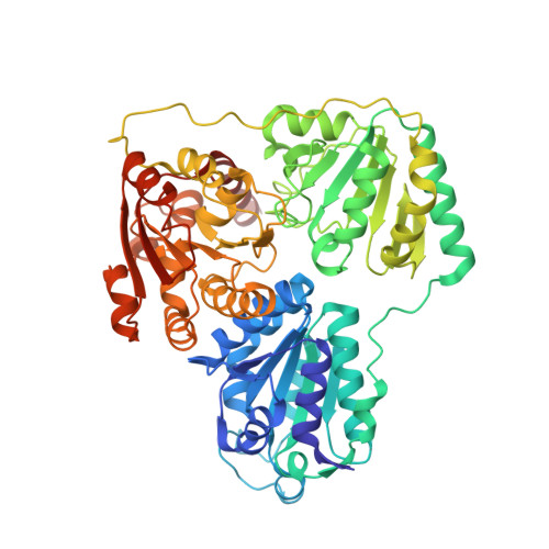
The mechanism by which the enzyme pyruvate decarboxylase from two yeast species is activated allosterically has been elucidated. A total of seven three-dimensional structures of the enzyme, of enzyme variants, or of enzyme complexes from two yeast species, three of them reported here for the first time, provide detailed atomic resolution snapshots along the activation coordinate. The prime event is the covalent binding of the substrate pyruvate to the side chain of cysteine 221, thus forming a thiohemiketal. This reaction causes the shift of a neighboring amino acid, which eventually leads to the rigidification of two otherwise flexible loops, one of which provides two histidine residues necessary to complete the enzymatically competent active site architecture. The structural data are complemented and supported by kinetic investigations and binding studies, providing a consistent picture of the structural changes occurring upon enzyme activation.
- Institute for Biochemistry and Biotechnology, Faculty of Biological Sciences, Martin-Luther-University Halle-Wittenberg, Kurt-Mothes-Str. 3, 06120 Halle (Saale), Germany.
Organizational Affiliation: