2UUE | pdb_00002uue

REPLACE: A strategy for Iterative Design of Cyclin Binding Groove Inhibitors


Experimental Data Snapshot

  • Method: X-RAY DIFFRACTION
  • Resolution: 2.06 Å
  • R-Value Free: 
    0.244 (Depositor), 0.244 (DCC) 
  • R-Value Work: 
    0.196 (Depositor), 0.199 (DCC) 
  • R-Value Observed: 
    0.197 (Depositor) 

Starting Model: experimental
View more details

wwPDB Validation 3D Report Full Report

Validation slider image for 2UUE

Ligand Structure Quality Assessment 


This is version 1.3 of the entry. See complete history

Literature

Replace: A Strategy for Iterative Design of Cyclin- Binding Groove Inhibitors

Andrews, M.J.Kontopidis, G.Mcinnes, C.Plater, A.Innes, L.Cowan, A.Jewsbury, P.Fischer, P.M.

(2006) Chembiochem 7: 1909

  • DOI: https://doi.org/10.1002/cbic.200600189
  • Primary Citation Related Structures: 
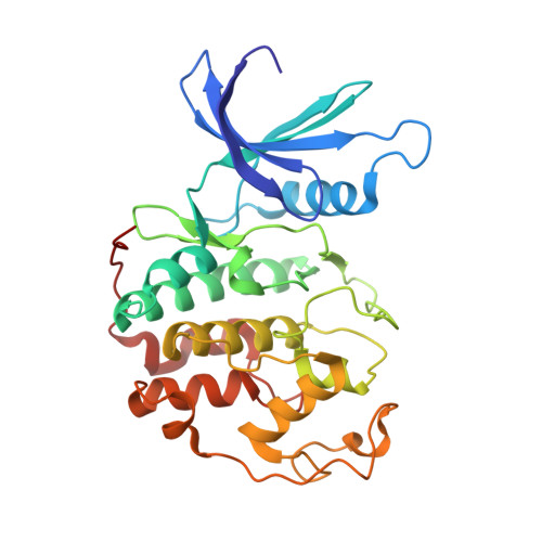
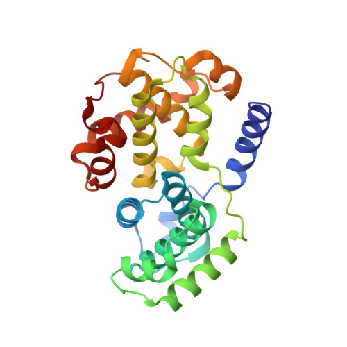
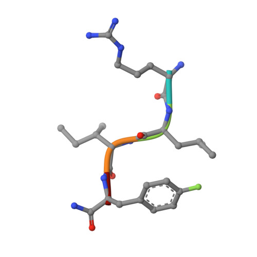
    2UUE, 2V22

  • PubMed Abstract: 

    We describe a drug-design strategy termed REPLACE (REplacement with Partial Ligand Alternatives through Computational Enrichment) in which nonpeptidic surrogates for specific determinants of known peptide ligands are identified in silico by using a core peptide-bound protein structure as a design anchor. In the REPLACE application example, we present the effective replacement of two critical binding motifs in a lead protein-protein interaction inhibitor pentapeptide with more druglike phenyltriazole and diphenyl ether groups. These were identified through docking of fragment libraries into the volume of the cyclin-binding groove of CDK2/cyclin A vacated through truncation of the inhibitor peptide-binding determinants. Proof of concept for this strategy was obtained through the generation of potent peptide-small-molecule hybrids and by the confirmation of inhibitor-binding modes in X-ray crystal structures. This method therefore allows nonpeptide fragments to be identified without the requirement for a high-sensitivity binding assay and should be generally applicable in replacing amino acids as individual residues or groups in peptide inhibitors to generate pharmaceutically acceptable lead molecules.


  • Organizational Affiliation
    • Cyclacel Limited, James Lindsay Place, Dundee, DD1 5JJ, UK. martin.andrews@glpg.com

Macromolecule Content 

  • Total Structure Weight: 129.87 kDa 
  • Atom Count: 9,332 
  • Modeled Residue Count: 1,119 
  • Deposited Residue Count: 1,124 
  • Unique protein chains: 3

Macromolecules

Find similar proteins by:|  3D Structure
Entity ID: 1
MoleculeChains  Sequence LengthOrganismDetailsImage
CELL DIVISION PROTEIN KINASE 2
A, C
298Homo sapiensMutation(s): 0 
EC: 2.7.11.1 (PDB Primary Data), 2.7.11.22 (UniProt)
UniProt & NIH Common Fund Data Resources
Find proteins for P24941 (Homo sapiens)
Explore P24941 
Go to UniProtKB:  P24941
PHAROS:  P24941
GTEx:  ENSG00000123374 
Entity Groups
Sequence Clusters30% Identity50% Identity70% Identity90% Identity95% Identity100% Identity
UniProt GroupP24941
Sequence Annotations
Expand
Reference Sequence
Find similar proteins by:|  3D Structure
Entity ID: 2
MoleculeChains  Sequence LengthOrganismDetailsImage
CYCLIN A2
B, D
259Homo sapiensMutation(s): 0 
UniProt & NIH Common Fund Data Resources
Find proteins for P20248 (Homo sapiens)
Explore P20248 
Go to UniProtKB:  P20248
PHAROS:  P20248
GTEx:  ENSG00000145386 
Entity Groups
Sequence Clusters30% Identity50% Identity70% Identity90% Identity95% Identity100% Identity
UniProt GroupP20248
Sequence Annotations
Expand
Reference Sequence
Find similar proteins by:  Sequence   |   3D Structure  
Entity ID: 3
MoleculeChains  Sequence LengthOrganismDetailsImage
GVC-TETRAPEPTIDE INHIBITOR
E, F
5synthetic constructMutation(s): 0 

Small Molecules

Modified Residues  1 Unique
IDChains TypeFormula2D DiagramParent
PFF
Query on PFF
E, F
L-PEPTIDE LINKINGC9 H10 F N O2PHE

Experimental Data & Validation

Experimental Data

  • Method: X-RAY DIFFRACTION
  • Resolution: 2.06 Å
  • R-Value Free:  0.244 (Depositor), 0.244 (DCC) 
  • R-Value Work:  0.196 (Depositor), 0.199 (DCC) 
  • R-Value Observed: 0.197 (Depositor) 
Space Group: P 21 21 21
Unit Cell:
Length ( Å )Angle ( ˚ )
a = 74.563α = 90
b = 113.8β = 90
c = 155.031γ = 90
Software Package:
Software NamePurpose
REFMACrefinement
MOSFLMdata reduction
SCALAdata scaling
MOLREPphasing

Structure Validation

View Full Validation Report


Ligand Structure Quality Assessment 


Entry History 

Deposition Data

Revision History  (Full details and data files)

  • Version 1.0: 2007-03-27
    Type: Initial release
  • Version 1.1: 2012-09-05
    Changes: Derived calculations, Non-polymer description, Other, Version format compliance
  • Version 1.2: 2019-10-09
    Changes: Data collection, Derived calculations, Other, Source and taxonomy
  • Version 1.3: 2023-12-13
    Changes: Data collection, Database references, Derived calculations, Refinement description