2RUS | pdb_00002rus

CRYSTAL STRUCTURE OF THE TERNARY COMPLEX OF RIBULOSE-1,5-BISPHOSPHATE CARBOXYLASE, MG(II), AND ACTIVATOR CO2 AT 2.3-ANGSTROMS RESOLUTION


Experimental Data Snapshot

  • Method: X-RAY DIFFRACTION
  • Resolution: 2.30 Å
  • R-Value Observed: 
    0.193 (Depositor) 

wwPDB Validation 3D Report Full Report

Validation slider image for 2RUS

This is version 1.4 of the entry. See complete history

Literature

Crystal structure of the ternary complex of ribulose-1,5-bisphosphate carboxylase, Mg(II), and activator CO2 at 2.3-A resolution.

Lundqvist, T.Schneider, G.

(1991) Biochemistry 30: 904-908

  • DOI: https://doi.org/10.1021/bi00218a004
  • Primary Citation Related Structures: 
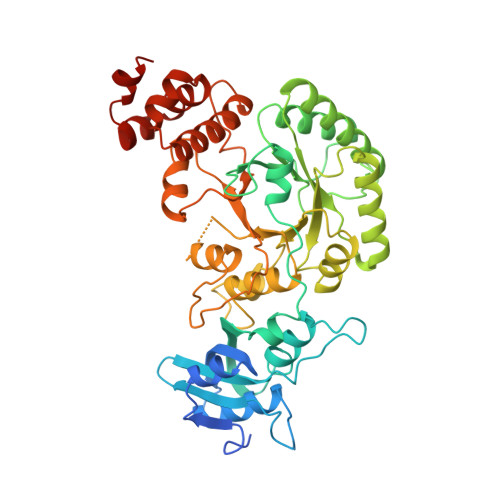
    2RUS

  • PubMed Abstract: 

    The activated ternary complex, enzyme-CO2-Mg(II), of the dimeric ribulose-1,5-bisphosphate carboxylase/oxygenase from Rhodospirillum rubrum can be prepared in the same crystal form that was used for the crystallographic structure determination of the native nonactivated enzyme (Schneider, G., Bränden, C.-I., & Lorimer, G. (1986) J. Mol. Biol. 187, 141-143). The three-dimensional structure of the activated enzyme has been determined to a nominal resolution of 2.3 A by protein crystallographic methods. The activator CO2 forms a carbamate with Lys191, located at the bottom of the funnel-shaped active site. In both subunits, this labile adduct is stabilized by a Mg(II) ion, bound to the carbamate and the side chains of Asp193 and Glu194. One solvent molecule was found within the first coordination sphere of the metal ion. The metal-binding site in ribulose-1,5-bisphosphate carboxylase consists thus of at least three protein ligands, all located on loop 2 of the beta/alpha barrel. One additional metal ligand, the side chain of the conserved Asn111, was observed close to the Mg(II) ion in the B-subunit. Other structural differences at the active site between the activated and nonactivated enzyme are limited to side-chain positions. Nevertheless, it is obvious that the hydrogen-bonding pattern in the vicinity of the activator site is completely altered.


  • Organizational Affiliation
    • Department of Molecular Biology, Swedish University of Agricultural Sciences, Uppsala.

Macromolecule Content 

  • Total Structure Weight: 106.65 kDa 
  • Atom Count: 7,445 
  • Modeled Residue Count: 881 
  • Deposited Residue Count: 980 
  • Unique protein chains: 1

Macromolecules

Find similar proteins by:|  3D Structure
Entity ID: 1
MoleculeChains  Sequence LengthOrganismDetailsImage
RUBISCO (RIBULOSE-1,5-BISPHOSPHATE CARBOXYLASE(SLASH)OXYGENASE)
A, B
490Rhodospirillum rubrumMutation(s): 0 
EC: 4.1.1.39
UniProt
Find proteins for P04718 (Rhodospirillum rubrum)
Explore P04718 
Go to UniProtKB:  P04718
Entity Groups
Sequence Clusters30% Identity50% Identity70% Identity90% Identity95% Identity100% Identity
UniProt GroupP04718
Sequence Annotations
Expand
Reference Sequence

Experimental Data & Validation

Experimental Data

  • Method: X-RAY DIFFRACTION
  • Resolution: 2.30 Å
  • R-Value Observed: 0.193 (Depositor) 
Space Group: P 1 21 1
Unit Cell:
Length ( Å )Angle ( ˚ )
a = 65.5α = 90
b = 70.6β = 92.1
c = 104.1γ = 90
Software Package:
Software NamePurpose
PROLSQrefinement

Structure Validation

View Full Validation Report


Entry History 

Deposition Data

Revision History  (Full details and data files)

  • Version 1.0: 1991-10-15
    Type: Initial release
  • Version 1.1: 2008-03-03
    Changes: Version format compliance
  • Version 1.2: 2011-07-13
    Changes: Version format compliance
  • Version 1.3: 2017-11-29
    Changes: Advisory, Derived calculations, Other
  • Version 1.4: 2025-03-26
    Changes: Advisory, Data collection, Database references, Derived calculations, Structure summary