2PHL

THE STRUCTURE OF PHASEOLIN AT 2.2 ANGSTROMS RESOLUTION: IMPLICATIONS FOR A COMMON VICILIN(SLASH)LEGUMIN STRUCTURE AND THE GENETIC ENGINEERING OF SEED STORAGE PROTEINS


Experimental Data Snapshot

  • Method: X-RAY DIFFRACTION
  • Resolution: 2.20 Å
  • R-Value Work: 0.178 
  • R-Value Observed: 0.178 

wwPDB Validation   3D Report Full Report


Ligand Structure Quality Assessment 


This is version 1.3 of the entry. See complete history


Literature

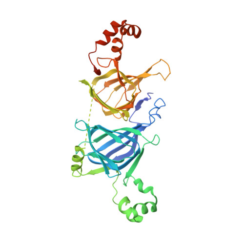
Structure of phaseolin at 2.2 A resolution. Implications for a common vicilin/legumin structure and the genetic engineering of seed storage proteins.

Lawrence, M.C.Izard, T.Beuchat, M.Blagrove, R.J.Colman, P.M.

(1994) J Mol Biol 238: 748-776

  • DOI: https://doi.org/10.1006/jmbi.1994.1333
  • Primary Citation of Related Structures:  
    2PHL

  • PubMed Abstract: 

    The refinement to 2.2 A resolution of the three-dimensional structure of the seed storage protein phaseolin from the French bean (Phaseolus vulgaris) via an alternative crystal form is described. The refined structure reveals details of the molecule hitherto unobserved and in particular we identify the structural role of conserved residues within the broader 7 S (vicilin) family of seed storage proteins. On this basis we are able to postulate a canonical model for the structure of the 7 S proteins. This model in turn provides a means for interpreting the structure of the 11 S (legumin) family of seed storage proteins, for which no X-ray diffraction data are available. The 11 S proteins are shown to bear a much closer relationship to the 7 S proteins than was previously recognized. The canonical model of the 7 S protein structure also provides a basis for proposing engineered mutations of these proteins with the goal of enhancing nutritional and functional properties.


  • Organizational Affiliation

    Biomolecular Research Institute, Parkville, Victoria, Australia.


Macromolecules
Find similar proteins by:  (by identity cutoff)  |  3D Structure
Entity ID: 1
MoleculeChains Sequence LengthOrganismDetailsImage
PHASEOLIN
A, B, C
397Phaseolus vulgarisMutation(s): 0 
UniProt
Find proteins for P02853 (Phaseolus vulgaris)
Explore P02853 
Go to UniProtKB:  P02853
Entity Groups  
Sequence Clusters30% Identity50% Identity70% Identity90% Identity95% Identity100% Identity
UniProt GroupP02853
Sequence Annotations
Expand
  • Reference Sequence
Experimental Data & Validation

Experimental Data

  • Method: X-RAY DIFFRACTION
  • Resolution: 2.20 Å
  • R-Value Work: 0.178 
  • R-Value Observed: 0.178 
  • Space Group: P 21 21 21
Unit Cell:
Length ( Å )Angle ( ˚ )
a = 89.81α = 90
b = 114.08β = 90
c = 137.08γ = 90
Software Package:
Software NamePurpose
X-PLORrefinement

Structure Validation

View Full Validation Report


Ligand Structure Quality Assessment 


Entry History 

Deposition Data

Revision History  (Full details and data files)

  • Version 1.0: 1994-09-30
    Type: Initial release
  • Version 1.1: 2008-03-24
    Changes: Version format compliance
  • Version 1.2: 2011-07-13
    Changes: Non-polymer description, Version format compliance
  • Version 1.3: 2020-07-29
    Type: Remediation
    Reason: Carbohydrate remediation
    Changes: Advisory, Data collection, Derived calculations, Other, Structure summary