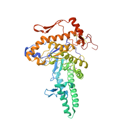
2PE4

Structure of Human Hyaluronidase 1, a Hyaluronan Hydrolyzing Enzyme Involved in Tumor Growth and Angiogenesis


Experimental Data Snapshot

  • Method: X-RAY DIFFRACTION
  • Resolution: 2.00 Å
  • R-Value Free: 0.230 
  • R-Value Work: 0.190 
  • R-Value Observed: 0.190 

Starting Model: experimental
View more details

wwPDB Validation   3D Report Full Report


This is version 2.1 of the entry. See complete history


Literature

Structure of Human Hyaluronidase-1, a Hyaluronan Hydrolyzing Enzyme Involved in Tumor Growth and Angiogenesis

Chao, K.L.Muthukumar, L.Herzberg, O.

(2007) Biochemistry 46: 6911-6920

  • DOI: https://doi.org/10.1021/bi700382g
  • Primary Citation of Related Structures:  
    2PE4

  • PubMed Abstract: 

    Mammalian hyaluronidases hydrolyze hyaluronan, a polysaccharide of diverse physiological roles found in all tissues and body fluids. In addition to its function in normal cellular hyaluronan turnover, human hyaluronidase-1 is implicated in cancer proliferation, angiogenesis, and inflammatory diseases; its expression is up-regulated in advanced stages of bladder cancer, whereas the expression of the alternative splice-variants is down-regulated. The crystal structure reveals a molecule composed of two closely associated domains: a catalytic domain that adopts a distorted (beta/alpha)8 barrel resembling that of bee venom hyaluronidase, and a novel, EGF-like domain, characteristic of involvement in protein-protein interactions and regulatory processes. The structure shows that the fold of this unique EGF-like domain is intact in four alternative splice-variants, whereas the catalytic domain is likely to be unfolded. Thus, these variants may function by competing with the full-length enzyme for the putative protein partner and regulating enzymatic activity in healthy cells.


  • Organizational Affiliation

    Center for Advanced Research in Biotechnology, University of Maryland Biotechnology Institute, Rockville, Maryland 20850, USA.


Macromolecules
Find similar proteins by:  (by identity cutoff)  |  3D Structure
Entity ID: 1
MoleculeChains Sequence LengthOrganismDetailsImage
Hyaluronidase-1424Homo sapiensMutation(s): 0 
Gene Names: HYAL1LUCA1
EC: 3.2.1.35
UniProt & NIH Common Fund Data Resources
Find proteins for Q12794 (Homo sapiens)
Explore Q12794 
Go to UniProtKB:  Q12794
PHAROS:  Q12794
GTEx:  ENSG00000114378 
Entity Groups  
Sequence Clusters30% Identity50% Identity70% Identity90% Identity95% Identity100% Identity
UniProt GroupQ12794
Glycosylation
Glycosylation Sites: 2Go to GlyGen: Q12794-1
Sequence Annotations
Expand
  • Reference Sequence
Oligosaccharides

Help

Entity ID: 2
MoleculeChains Length2D Diagram Glycosylation3D Interactions
alpha-D-mannopyranose-(1-3)-alpha-D-mannopyranose-(1-6)-[alpha-D-mannopyranose-(1-3)]beta-D-mannopyranose-(1-4)-2-acetamido-2-deoxy-beta-D-glucopyranose-(1-4)-2-acetamido-2-deoxy-beta-D-glucopyranose
B
6N-Glycosylation
Glycosylation Resources
GlyTouCan:  G09724ZC
GlyCosmos:  G09724ZC
GlyGen:  G09724ZC
Entity ID: 3
MoleculeChains Length2D Diagram Glycosylation3D Interactions
2-acetamido-2-deoxy-beta-D-glucopyranose-(1-4)-2-acetamido-2-deoxy-beta-D-glucopyranose
C
2N-Glycosylation
Glycosylation Resources
GlyTouCan:  G42666HT
GlyCosmos:  G42666HT
GlyGen:  G42666HT
Experimental Data & Validation

Experimental Data

  • Method: X-RAY DIFFRACTION
  • Resolution: 2.00 Å
  • R-Value Free: 0.230 
  • R-Value Work: 0.190 
  • R-Value Observed: 0.190 
  • Space Group: P 32 2 1
Unit Cell:
Length ( Å )Angle ( ˚ )
a = 92.05α = 90
b = 92.05β = 90
c = 143.81γ = 120
Software Package:
Software NamePurpose
d*TREKdata scaling
PHASERphasing
REFMACrefinement
PDB_EXTRACTdata extraction
CrystalCleardata collection
d*TREKdata reduction

Structure Validation

View Full Validation Report


Entry History 

Deposition Data

Revision History  (Full details and data files)

  • Version 1.0: 2007-06-12
    Type: Initial release
  • Version 1.1: 2008-05-01
    Changes: Version format compliance
  • Version 1.2: 2011-07-13
    Changes: Non-polymer description, Version format compliance
  • Version 1.3: 2017-10-18
    Changes: Refinement description
  • Version 2.0: 2020-07-29
    Type: Remediation
    Reason: Carbohydrate remediation
    Changes: Atomic model, Data collection, Database references, Derived calculations, Structure summary
  • Version 2.1: 2023-08-30
    Changes: Data collection, Database references, Refinement description, Structure summary