2PAE

Structure of a H49N mutant dTDP-4-keto-6-deoxy-D-glucose-3,4-ketoisomerase from Aneurinibacillus thermoaerophilus in complex with TDP


Experimental Data Snapshot

  • Method: X-RAY DIFFRACTION
  • Resolution: 2.50 Å
  • R-Value Free: 0.256 
  • R-Value Work: 0.207 
  • R-Value Observed: 0.200 

wwPDB Validation   3D Report Full Report


Ligand Structure Quality Assessment 


This is version 1.4 of the entry. See complete history


Literature

The X-ray Structure of dTDP-4-Keto-6-deoxy-D-glucose-3,4-ketoisomerase.

Davis, M.L.Thoden, J.B.Holden, H.M.

(2007) J Biol Chem 282: 19227-19236

  • DOI: https://doi.org/10.1074/jbc.M702529200
  • Primary Citation of Related Structures:  
    2PA7, 2PAE, 2PAK, 2PAM

  • PubMed Abstract: 

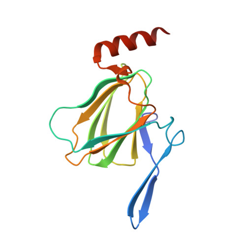
    The repeating unit of the glycan chain in the S-layer of the bacterium Aneurinibacillus thermoaerophilus L420-91(T) is composed of four alpha-d-rhamnose molecules and two 3-acetamido-3,6-dideoxy-alpha-d-galactose moieties (abbreviated as Fucp3NAc). Formation of the glycan layer requires nucleotide-activated sugars as the donor molecules. Whereas the enzymes involved in the synthesis of GDP-rhamnose have been well characterized, less is known regarding the structures and enzymatic mechanisms of the enzymes required for the production of dTDP-Fucp3NAc. One of the enzymes involved in the biosynthesis of dTDP-Fucp3NAc is a 3,4-ketoisomerase, hereafter referred to as FdtA. Here we describe the first three-dimensional structure of this sugar isomerase complexed with dTDP and solved to 1.5 A resolution. The FdtA dimer assumes an almost jellyfish-like appearance with the sole alpha-helices representing the tentacles. Formation of the FdtA dimer represents a classical example of domain swapping whereby beta-strands 2 and 3 from one subunit form part of a beta-sheet in the second subunit. The active site architecture of FdtA is characterized by a cluster of three histidine residues, two of which, His(49) and His(51), appear to be strictly conserved in the amino acid sequences deposited to date. Site-directed mutagenesis experiments, enzymatic assays, and x-ray crystallographic analyses suggest that His(49) functions as an active site base.


  • Organizational Affiliation

    Department of Biochemistry, University of Wisconsin, Madison, Wisconsin 53706-1544, USA.


Macromolecules
Find similar proteins by:  (by identity cutoff)  |  3D Structure
Entity ID: 1
MoleculeChains Sequence LengthOrganismDetailsImage
DTDP-6-deoxy-3,4-keto-hexulose isomerase
A, B
141Aneurinibacillus thermoaerophilusMutation(s): 1 
Gene Names: fdtA
EC: 5.3.1
UniProt
Find proteins for Q6T1W8 (Aneurinibacillus thermoaerophilus)
Explore Q6T1W8 
Go to UniProtKB:  Q6T1W8
Entity Groups  
Sequence Clusters30% Identity50% Identity70% Identity90% Identity95% Identity100% Identity
UniProt GroupQ6T1W8
Sequence Annotations
Expand
  • Reference Sequence
Small Molecules
Experimental Data & Validation

Experimental Data

  • Method: X-RAY DIFFRACTION
  • Resolution: 2.50 Å
  • R-Value Free: 0.256 
  • R-Value Work: 0.207 
  • R-Value Observed: 0.200 
  • Space Group: P 41 21 2
Unit Cell:
Length ( Å )Angle ( ˚ )
a = 61.6α = 90
b = 61.6β = 90
c = 202.2γ = 90
Software Package:
Software NamePurpose
PROTEUM PLUSdata collection
EPMRphasing
TNTrefinement
SAINTdata reduction
SADABSdata scaling

Structure Validation

View Full Validation Report


Ligand Structure Quality Assessment 


Entry History 

Deposition Data

Revision History  (Full details and data files)

  • Version 1.0: 2007-04-24
    Type: Initial release
  • Version 1.1: 2008-05-01
    Changes: Version format compliance
  • Version 1.2: 2011-07-13
    Changes: Version format compliance
  • Version 1.3: 2021-10-20
    Changes: Database references, Derived calculations
  • Version 1.4: 2023-08-30
    Changes: Data collection, Refinement description