2NT8

ATP bound at the active site of a PduO type ATP:co(I)rrinoid adenosyltransferase from Lactobacillus reuteri


Experimental Data Snapshot

  • Method: X-RAY DIFFRACTION
  • Resolution: 1.68 Å
  • R-Value Free: 0.204 
  • R-Value Work: 0.173 
  • R-Value Observed: 0.175 

wwPDB Validation   3D Report Full Report


Ligand Structure Quality Assessment 


This is version 1.4 of the entry. See complete history


Literature

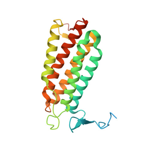
Structural characterization of the active site of the PduO-type ATP:Co(I)rrinoid adenosyltransferase from Lactobacillus reuteri.

St Maurice, M.Mera, P.E.Taranto, M.P.Sesma, F.Escalante-Semerena, J.C.Rayment, I.

(2007) J Biol Chem 282: 2596-2605

  • DOI: https://doi.org/10.1074/jbc.M609557200
  • Primary Citation of Related Structures:  
    2NT8

  • PubMed Abstract: 

    The three-dimensional crystal structure of the PduO-type corrinoid adenosyltransferase from Lactobacillus reuteri (LrPduO) has been solved to 1.68-A resolution. The functional assignment of LrPduO as a corrinoid adenosyltransferase was confirmed by in vivo and in vitro evidence. The enzyme has an apparent Km(ATP) of 2.2 microM and Km(Cobalamin) of 0.13 microM and a kcat of 0.025 s(-1). Co-crystallization of the enzyme with Mg-ATP resulted in well-defined electron density for an N-terminal loop that had been disordered in other PduO-type enzyme structures. This newly defined N-terminal loop makes up the lower portion of the enzyme active site with the other half being contributed from an adjacent subunit. These results provide the first detailed description of the enzyme active site for a PduO-type adenosyltransferase and identify a unique ATP binding motif at the protein N terminus. The molecular architecture at the active site offers valuable new insight into the role of various residues responsible for the human disease methylmalonic aciduria.


  • Organizational Affiliation

    Department of Biochemistry, University of Wisconsin, Madison, Wisconsin 53706, USA.


Macromolecules
Find similar proteins by:  (by identity cutoff)  |  3D Structure
Entity ID: 1
MoleculeChains Sequence LengthOrganismDetailsImage
Cobalamin adenosyltransferase223Limosilactobacillus reuteriMutation(s): 0 
Gene Names: pduo
EC: 2.5.1.17
UniProt
Find proteins for Q50EJ2 (Limosilactobacillus reuteri)
Explore Q50EJ2 
Go to UniProtKB:  Q50EJ2
Entity Groups  
Sequence Clusters30% Identity50% Identity70% Identity90% Identity95% Identity100% Identity
UniProt GroupQ50EJ2
Sequence Annotations
Expand
  • Reference Sequence
Experimental Data & Validation

Experimental Data

  • Method: X-RAY DIFFRACTION
  • Resolution: 1.68 Å
  • R-Value Free: 0.204 
  • R-Value Work: 0.173 
  • R-Value Observed: 0.175 
  • Space Group: I 2 3
Unit Cell:
Length ( Å )Angle ( ˚ )
a = 110.609α = 90
b = 110.609β = 90
c = 110.609γ = 90
Software Package:
Software NamePurpose
DENZOdata reduction
SCALEPACKdata scaling
REFMACrefinement
PDB_EXTRACTdata extraction
HKL-2000data reduction
HKL-2000data scaling

Structure Validation

View Full Validation Report


Ligand Structure Quality Assessment 


Entry History 

Deposition Data

Revision History  (Full details and data files)

  • Version 1.0: 2006-11-21
    Type: Initial release
  • Version 1.1: 2008-05-01
    Changes: Version format compliance
  • Version 1.2: 2011-07-13
    Changes: Derived calculations, Version format compliance
  • Version 1.3: 2017-10-18
    Changes: Refinement description
  • Version 1.4: 2023-08-30
    Changes: Data collection, Database references, Derived calculations, Refinement description