2JXY | pdb_00002jxy

Solution structure of the hemopexin-like domain of MMP12


Experimental Data Snapshot

  • Method: SOLUTION NMR
  • Conformers Calculated: 1600 
  • Conformers Submitted: 20 
  • Selection Criteria: lowest target function 

wwPDB Validation 3D Report Full Report

Validation slider image for 2JXY

This is version 1.3 of the entry. See complete history

Literature

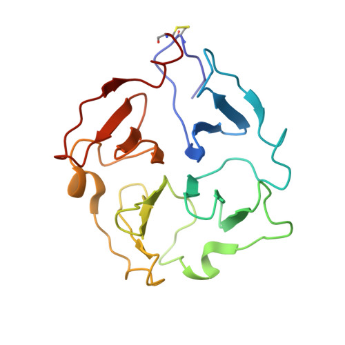
Evidence of reciprocal reorientation of the catalytic and hemopexin-like domains of full-length MMP-12

Bertini, I.Calderone, V.Fragai, M.Jaiswal, R.Luchinat, C.Melikian, M.Mylonas, E.Svergun, D.I.

(2008) J Am Chem Soc 130: 7011-7021

  • DOI: https://doi.org/10.1021/ja710491y
  • Primary Citation Related Structures: 
    2JXY, 3BA0

  • PubMed Abstract: 

    The proteolytic activity of matrix metalloproteinases toward extracellular matrix components (ECM), cytokines, chemokines, and membrane receptors is crucial for several homeostatic and pathological processes. Active MMPs are a family of single-chain enzymes (23 family members in the human genome), most of which constituted by a catalytic domain and by a hemopexin-like domain connected by a linker. The X-ray structures of MMP-1 and MMP-2 suggest a conserved and well-defined spatial relationship between the two domains. Here we present structural data for MMP-12, suitably stabilized against self-hydrolysis, both in solution (NMR and SAXS) and in the solid state (X-ray), showing that the hemopexin-like and the catalytic domains experience conformational freedom with respect to each other on a time scale shorter than 10(-8) s. Hints on the probable conformations are also obtained. This experimental finding opens new perspectives for the often hypothesized active role of the hemopexin-like domain in the enzymatic activity of MMPs.


  • Organizational Affiliation
    • Magnetic Resonance Center (CERM), University of Florence, Via L. Sacconi 6, 50019 Sesto Fiorentino, Italy. ivanobertini@cerm.unifi.it

Macromolecule Content 

  • Total Structure Weight: 23.24 kDa 
  • Atom Count: 1,648 
  • Modeled Residue Count: 194 
  • Deposited Residue Count: 194 
  • Unique protein chains: 1

Macromolecules

Find similar proteins by:|  3D Structure
Entity ID: 1
MoleculeChains  Sequence LengthOrganismDetailsImage
Macrophage metalloelastase194Homo sapiensMutation(s): 0 
EC: 3.4.24.65
UniProt & NIH Common Fund Data Resources
Find proteins for P39900 (Homo sapiens)
Explore P39900 
Go to UniProtKB:  P39900
PHAROS:  P39900
GTEx:  ENSG00000262406 
Entity Groups
Sequence Clusters30% Identity50% Identity70% Identity90% Identity95% Identity100% Identity
UniProt GroupP39900
Sequence Annotations
Expand
Reference Sequence

Small Molecules

Ligands 1 Unique
IDChains Name / Formula / InChI Key2D Diagram3D Interactions
CA

Query on CA


Download:Ideal Coordinates CCD File
B [auth A]CALCIUM ION
Ca
BHPQYMZQTOCNFJ-UHFFFAOYSA-N

Experimental Data & Validation

Experimental Data

  • Method: SOLUTION NMR
  • Conformers Calculated: 1600 
  • Conformers Submitted: 20 
  • Selection Criteria: lowest target function 

Structure Validation

View Full Validation Report


Entry History 

Deposition Data

Revision History  (Full details and data files)

  • Version 1.0: 2008-05-27
    Type: Initial release
  • Version 1.1: 2011-07-13
    Changes: Version format compliance
  • Version 1.2: 2022-03-16
    Changes: Data collection, Database references, Derived calculations
  • Version 1.3: 2024-11-13
    Changes: Data collection, Structure summary