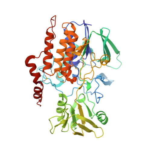
2GVC

Crystal structure of flavin-containing monooxygenase (FMO)from S.pombe and substrate (methimazole) complex


Experimental Data Snapshot

  • Method: X-RAY DIFFRACTION
  • Resolution: 2.22 Å
  • R-Value Free: 0.265 
  • R-Value Work: 0.230 
  • R-Value Observed: 0.230 

Starting Model: experimental
View more details

wwPDB Validation   3D Report Full Report


Ligand Structure Quality Assessment 


This is version 1.5 of the entry. See complete history


Literature

Mechanism of action of a flavin-containing monooxygenase.

Eswaramoorthy, S.Bonanno, J.B.Burley, S.K.Swaminathan, S.

(2006) Proc Natl Acad Sci U S A 103: 9832-9837

  • DOI: https://doi.org/10.1073/pnas.0602398103
  • Primary Citation of Related Structures:  
    1VQW, 2GV8, 2GVC

  • PubMed Abstract: 

    Elimination of nonnutritional and insoluble compounds is a critical task for any living organism. Flavin-containing monooxygenases (FMOs) attach an oxygen atom to the insoluble nucleophilic compounds to increase solubility and thereby increase excretion. Here we analyze the functional mechanism of FMO from Schizosaccharomyces pombe using the crystal structures of the wild type and protein-cofactor and protein-substrate complexes. The structure of the wild-type FMO revealed that the prosthetic group FAD is an integral part of the protein. FMO needs NADPH as a cofactor in addition to the prosthetic group for its catalytic activity. Structures of the protein-cofactor and protein-substrate complexes provide insights into mechanism of action. We propose that FMOs exist in the cell as a complex with a reduced form of the prosthetic group and NADPH cofactor, readying them to act on substrates. The 4alpha-hydroperoxyflavin form of the prosthetic group represents a transient intermediate of the monooxygenation process. The oxygenated and reduced forms of the prosthetic group help stabilize interactions with cofactor and substrate alternately to permit continuous enzyme turnover.


  • Organizational Affiliation

    Biology Department, Brookhaven National Laboratory, Upton, NY 11973, USA.


Macromolecules
Find similar proteins by:  (by identity cutoff)  |  3D Structure
Entity ID: 1
MoleculeChains Sequence LengthOrganismDetailsImage
monooxygenaseA,
B,
C [auth D],
D [auth E]
447Schizosaccharomyces pombeMutation(s): 5 
UniProt
Find proteins for Q9HFE4 (Schizosaccharomyces pombe (strain 972 / ATCC 24843))
Explore Q9HFE4 
Go to UniProtKB:  Q9HFE4
Entity Groups  
Sequence Clusters30% Identity50% Identity70% Identity90% Identity95% Identity100% Identity
UniProt GroupQ9HFE4
Sequence Annotations
Expand
  • Reference Sequence
Small Molecules
Ligands 4 Unique
IDChains Name / Formula / InChI Key2D Diagram3D Interactions
FAD
Query on FAD

Download Ideal Coordinates CCD File 
F [auth A],
J [auth B],
N [auth D],
R [auth E]
FLAVIN-ADENINE DINUCLEOTIDE
C27 H33 N9 O15 P2
VWWQXMAJTJZDQX-UYBVJOGSSA-N
MMZ
Query on MMZ

Download Ideal Coordinates CCD File 
G [auth A],
K [auth B],
O [auth D],
S [auth E]
1-METHYL-1,3-DIHYDRO-2H-IMIDAZOLE-2-THIONE
C4 H6 N2 S
PMRYVIKBURPHAH-UHFFFAOYSA-N
GOL
Query on GOL

Download Ideal Coordinates CCD File 
H [auth A],
L [auth B],
P [auth D],
T [auth E]
GLYCEROL
C3 H8 O3
PEDCQBHIVMGVHV-UHFFFAOYSA-N
PEO
Query on PEO

Download Ideal Coordinates CCD File 
E [auth A],
I [auth B],
M [auth D],
Q [auth E]
HYDROGEN PEROXIDE
H2 O2
MHAJPDPJQMAIIY-UHFFFAOYSA-N
Modified Residues  1 Unique
IDChains TypeFormula2D DiagramParent
MSE
Query on MSE
A,
B,
C [auth D],
D [auth E]
L-PEPTIDE LINKINGC5 H11 N O2 SeMET
Experimental Data & Validation

Experimental Data

  • Method: X-RAY DIFFRACTION
  • Resolution: 2.22 Å
  • R-Value Free: 0.265 
  • R-Value Work: 0.230 
  • R-Value Observed: 0.230 
  • Space Group: P 1
Unit Cell:
Length ( Å )Angle ( ˚ )
a = 72.7α = 107.73
b = 84.45β = 90.76
c = 113.51γ = 106.5
Software Package:
Software NamePurpose
CBASSdata collection
SCALEPACKdata scaling
MOLREPphasing
CNSrefinement

Structure Validation

View Full Validation Report


Ligand Structure Quality Assessment 


Entry History 

Deposition Data

Revision History  (Full details and data files)

  • Version 1.0: 2006-06-06
    Type: Initial release
  • Version 1.1: 2008-05-01
    Changes: Version format compliance
  • Version 1.2: 2011-07-13
    Changes: Non-polymer description, Version format compliance
  • Version 1.3: 2021-02-03
    Changes: Database references, Derived calculations, Structure summary
  • Version 1.4: 2023-08-30
    Changes: Data collection, Database references, Refinement description
  • Version 1.5: 2023-11-15
    Changes: Data collection