2A1T

Structure of the human MCAD:ETF E165betaA complex


Experimental Data Snapshot

  • Method: X-RAY DIFFRACTION
  • Resolution: 2.80 Å
  • R-Value Free: 0.269 
  • R-Value Work: 0.199 
  • R-Value Observed: 0.203 

wwPDB Validation   3D Report Full Report


Ligand Structure Quality Assessment 


This is version 1.4 of the entry. See complete history


Literature

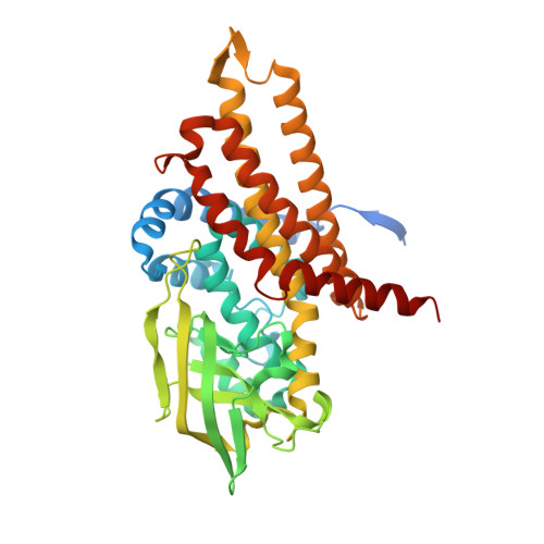
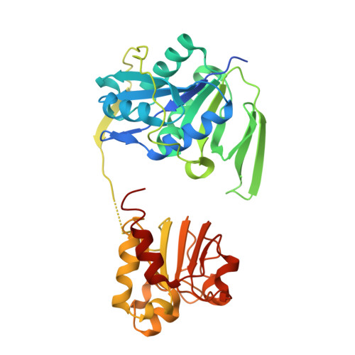
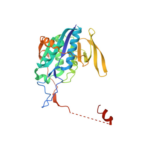
Stabilization of Non-productive Conformations Underpins Rapid Electron Transfer to Electron-transferring Flavoprotein

Toogood, H.S.van Thiel, A.Scrutton, N.S.Leys, D.

(2005) J Biol Chem 280: 30361-30366

  • DOI: https://doi.org/10.1074/jbc.M505562200
  • Primary Citation of Related Structures:  
    2A1T, 2A1U

  • PubMed Abstract: 
  • Crystal structures of protein complexes with electron-transferring flavoprotein (ETF) have revealed a dual protein-protein interface with one region serving as anchor while the ETF FAD domain samples available space within the complex. We show that mutation of the conserved Glu-165beta in human ETF leads to drastically modulated rates of interprotein electron transfer with both medium chain acyl-CoA dehydrogenase and dimethylglycine dehydrogenase ...

    Crystal structures of protein complexes with electron-transferring flavoprotein (ETF) have revealed a dual protein-protein interface with one region serving as anchor while the ETF FAD domain samples available space within the complex. We show that mutation of the conserved Glu-165beta in human ETF leads to drastically modulated rates of interprotein electron transfer with both medium chain acyl-CoA dehydrogenase and dimethylglycine dehydrogenase. The crystal structure of free E165betaA ETF is essentially identical to that of wild-type ETF, but the crystal structure of the E165betaA ETF.medium chain acyl-CoA dehydrogenase complex reveals clear electron density for the FAD domain in a position optimal for fast interprotein electron transfer. Based on our observations, we present a dynamic multistate model for conformational sampling that for the wild-type ETF. medium chain acyl-CoA dehydrogenase complex involves random motion between three distinct positions for the ETF FAD domain. ETF Glu-165beta plays a key role in stabilizing positions incompatible with fast interprotein electron transfer, thus ensuring high rates of complex dissociation.


    Organizational Affiliation

    Department of Biochemistry, University of Leicester, Henry Wellcome Building, Lancaster Road, LE1 7RH, Leicester United Kingdom.


Macromolecules
Find similar proteins by:  (by identity cutoff)  |  3D Structure
Entity ID: 1
MoleculeChainsSequence LengthOrganismDetailsImage
Acyl-CoA dehydrogenase, medium-chain specific, mitochondrial precursor
A, B, C, D
421Homo sapiensMutation(s): 0 
Gene Names: ACADM
EC: 1.3.99.3 (PDB Primary Data), 1.3.8.7 (UniProt)
UniProt & NIH Common Fund Data Resources
Find proteins for P11310 (Homo sapiens)
Explore P11310 
Go to UniProtKB:  P11310
PHAROS:  P11310
Entity Groups  
Sequence Clusters30% Identity50% Identity70% Identity90% Identity95% Identity100% Identity
UniProt GroupP11310
Protein Feature View
Expand
  • Reference Sequence
Find similar proteins by:  (by identity cutoff)  |  3D Structure
Entity ID: 2
MoleculeChainsSequence LengthOrganismDetailsImage
Electron transfer flavoprotein alpha-subunit, mitochondrial precursorE [auth R]333Homo sapiensMutation(s): 0 
Gene Names: ETFA
UniProt & NIH Common Fund Data Resources
Find proteins for P13804 (Homo sapiens)
Explore P13804 
Go to UniProtKB:  P13804
PHAROS:  P13804
Entity Groups  
Sequence Clusters30% Identity50% Identity70% Identity90% Identity95% Identity100% Identity
UniProt GroupP13804
Protein Feature View
Expand
  • Reference Sequence
Find similar proteins by:  (by identity cutoff)  |  3D Structure
Entity ID: 3
MoleculeChainsSequence LengthOrganismDetailsImage
Electron transfer flavoprotein beta-subunitF [auth S]255Homo sapiensMutation(s): 1 
Gene Names: ETFBFP585
UniProt & NIH Common Fund Data Resources
Find proteins for P38117 (Homo sapiens)
Explore P38117 
Go to UniProtKB:  P38117
PHAROS:  P38117
Entity Groups  
Sequence Clusters30% Identity50% Identity70% Identity90% Identity95% Identity100% Identity
UniProt GroupP38117
Protein Feature View
Expand
  • Reference Sequence
Small Molecules
Ligands 2 Unique
IDChainsName / Formula / InChI Key2D Diagram3D Interactions
FAD
Query on FAD

Download Ideal Coordinates CCD File 
G [auth A],
H [auth B],
I [auth C],
J [auth D],
K [auth R]
FLAVIN-ADENINE DINUCLEOTIDE
C27 H33 N9 O15 P2
VWWQXMAJTJZDQX-UYBVJOGSSA-N
 Ligand Interaction
AMP
Query on AMP

Download Ideal Coordinates CCD File 
L [auth S]ADENOSINE MONOPHOSPHATE
C10 H14 N5 O7 P
UDMBCSSLTHHNCD-KQYNXXCUSA-N
 Ligand Interaction
Experimental Data & Validation

Experimental Data

  • Method: X-RAY DIFFRACTION
  • Resolution: 2.80 Å
  • R-Value Free: 0.269 
  • R-Value Work: 0.199 
  • R-Value Observed: 0.203 
  • Space Group: P 21 21 21
Unit Cell:
Length ( Å )Angle ( ˚ )
a = 94.184α = 90
b = 100.659β = 90
c = 244.384γ = 90
Software Package:
Software NamePurpose
REFMACrefinement
HKL-2000data reduction
SCALEPACKdata scaling
AMoREphasing

Structure Validation

View Full Validation Report


Ligand Structure Quality Assessment 


Entry History 

Deposition Data

Revision History  (Full details and data files)

  • Version 1.0: 2005-07-05
    Type: Initial release
  • Version 1.1: 2008-04-30
    Changes: Version format compliance
  • Version 1.2: 2011-07-13
    Changes: Version format compliance
  • Version 1.3: 2018-10-03
    Changes: Data collection
  • Version 1.4: 2021-11-10
    Changes: Database references, Derived calculations