2ZXC | pdb_00002zxc

Ceramidase complexed with C2


Experimental Data Snapshot

  • Method: X-RAY DIFFRACTION
  • Resolution: 2.20 Å
  • R-Value Free: 
    0.228 (Depositor), 0.227 (DCC) 
  • R-Value Work: 
    0.194 (Depositor), 0.194 (DCC) 
  • R-Value Observed: 
    0.194 (Depositor) 

Starting Model: experimental
View more details

wwPDB Validation 3D Report Full Report

Validation slider image for 2ZXC

Ligand Structure Quality Assessment 


This is version 1.4 of the entry. See complete history

Literature

Mechanistic insights into the hydrolysis and synthesis of ceramide by neutral ceramidase.

Inoue, T.Okino, N.Kakuta, Y.Hijikata, A.Okano, H.Goda, H.M.Tani, M.Sueyoshi, N.Kambayashi, K.Matsumura, H.Kai, Y.Ito, M.

(2009) J Biological Chem 284: 9566-9577

  • DOI: https://doi.org/10.1074/jbc.M808232200
  • Primary Citation Related Structures: 
    2ZWS, 2ZXC

  • PubMed Abstract: 

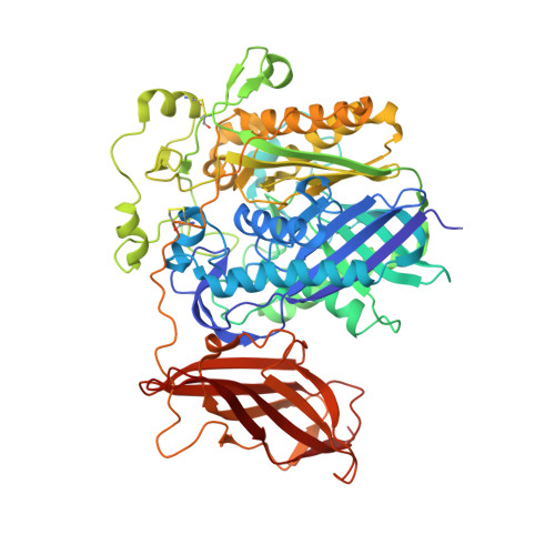
    Ceramidase (CDase; EC 3.5.1.23) hydrolyzes ceramide to generate sphingosine and fatty acid. The enzyme plays a regulatory role in a variety of physiological events in eukaryotes and also functions as an exotoxin in particular bacteria. The crystal structures of neutral CDase from Pseudomonas aeruginosa (PaCD) in the C2-ceramide-bound and -unbound forms were determined at 2.2 and 1.4 A resolutions, respectively. PaCD consists of two domains, and the Zn(2+)- and Mg(2+)/Ca(2+)-binding sites are found within the center of the N-terminal domain and the interface between the domains, respectively. The structural comparison between the C2-ceramide-bound and unbound forms revealed an open-closed conformational change occurring to loop I upon binding of C2-ceramide. In the closed state, this loop sits above the Zn(2+) coordination site and over the opening to the substrate binding site. Mutational analyses of residues surrounding the Zn(2+) of PaCD and rat neutral CDase revealed that the cleavage or creation of the N-acyl linkage of ceramide follows a similar mechanism as observed for the Zn(2+)-dependent carboxypeptidases. The results provide an understanding of the molecular mechanism of hydrolysis and synthesis of ceramide by the enzyme. Furthermore, insights into the actions of PaCD and eukaryotic neutral CDases as an exotoxin and mediators of sphingolipid signaling are also revealed, respectively.


  • Organizational Affiliation
    • Department of Applied Chemistry, Graduate School of Engineering, Osaka University, 2-1 Yamada-Oka, Suita, Osaka 565-0871, Japan. inouet@chem.eng.osaka-u.ac.jp

Macromolecule Content 

  • Total Structure Weight: 143.1 kDa 
  • Atom Count: 10,943 
  • Modeled Residue Count: 1,286 
  • Deposited Residue Count: 1,292 
  • Unique protein chains: 1

Macromolecules

Find similar proteins by:|  3D Structure
Entity ID: 1
MoleculeChains  Sequence LengthOrganismDetailsImage
Neutral ceramidase
A, B
646Pseudomonas aeruginosaMutation(s): 0 
EC: 3.5.1.23
Membrane Entity: Yes 
UniProt
Find proteins for Q9I596 (Pseudomonas aeruginosa (strain ATCC 15692 / DSM 22644 / CIP 104116 / JCM 14847 / LMG 12228 / 1C / PRS 101 / PAO1))
Explore Q9I596 
Go to UniProtKB:  Q9I596
Entity Groups
Sequence Clusters30% Identity50% Identity70% Identity90% Identity95% Identity100% Identity
UniProt GroupQ9I596
Sequence Annotations
Expand
Reference Sequence

Small Molecules

Ligands 5 Unique
IDChains Name / Formula / InChI Key2D Diagram3D Interactions
2ED

Query on 2ED


Download:Ideal Coordinates CCD File
D [auth A],
K [auth B]
N-[(1R,2R,3E)-2-hydroxy-1-(hydroxymethyl)heptadec-3-en-1-yl]acetamide
C20 H39 N O3
BLTCBVOJNNKFKC-KTEGJIGUSA-N
DMS

Query on DMS


Download:Ideal Coordinates CCD File
P [auth B],
Q [auth B]
DIMETHYL SULFOXIDE
C2 H6 O S
IAZDPXIOMUYVGZ-UHFFFAOYSA-N
ZN

Query on ZN


Download:Ideal Coordinates CCD File
C [auth A],
J [auth B]
ZINC ION
Zn
PTFCDOFLOPIGGS-UHFFFAOYSA-N
FMT

Query on FMT


Download:Ideal Coordinates CCD File
E [auth A]
F [auth A]
G [auth A]
H [auth A]
L [auth B]
E [auth A],
F [auth A],
G [auth A],
H [auth A],
L [auth B],
M [auth B],
N [auth B],
O [auth B]
FORMIC ACID
C H2 O2
BDAGIHXWWSANSR-UHFFFAOYSA-N
MG

Query on MG


Download:Ideal Coordinates CCD File
I [auth A],
R [auth B]
MAGNESIUM ION
Mg
JLVVSXFLKOJNIY-UHFFFAOYSA-N

Experimental Data & Validation

Experimental Data

  • Method: X-RAY DIFFRACTION
  • Resolution: 2.20 Å
  • R-Value Free:  0.228 (Depositor), 0.227 (DCC) 
  • R-Value Work:  0.194 (Depositor), 0.194 (DCC) 
  • R-Value Observed: 0.194 (Depositor) 
Space Group: P 21 21 21
Unit Cell:
Length ( Å )Angle ( ˚ )
a = 65.741α = 90
b = 65.8β = 90
c = 340.339γ = 90
Software Package:
Software NamePurpose
HKL-2000data collection
MOLREPphasing
CNSrefinement
HKL-2000data reduction
HKL-2000data scaling

Structure Validation

View Full Validation Report


Ligand Structure Quality Assessment 


Entry History 

Deposition Data

Revision History  (Full details and data files)

  • Version 1.0: 2009-02-03
    Type: Initial release
  • Version 1.1: 2011-07-13
    Changes: Version format compliance
  • Version 1.2: 2017-10-04
    Changes: Structure summary
  • Version 1.3: 2024-04-03
    Changes: Data collection, Database references, Derived calculations, Refinement description
  • Version 1.4: 2024-10-23
    Changes: Structure summary