2ZNH

Crystal Structure of a Domain-Swapped Serpin Dimer


Experimental Data Snapshot

  • Method: X-RAY DIFFRACTION
  • Resolution: 2.80 Å
  • R-Value Free: 0.247 
  • R-Value Work: 0.201 
  • R-Value Observed: 0.201 

wwPDB Validation   3D Report Full Report


Ligand Structure Quality Assessment 


This is version 2.1 of the entry. See complete history


Literature

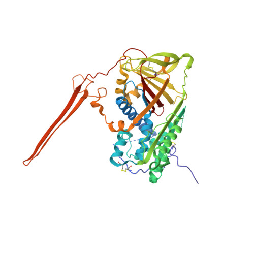
Crystal structure of a stable dimer reveals the molecular basis of serpin polymerization

Yamasaki, M.Li, W.Johnson, D.J.Huntington, J.A.

(2008) Nature 455: 1255-1258

  • DOI: https://doi.org/10.1038/nature07394
  • Primary Citation of Related Structures:  
    2ZNH

  • PubMed Abstract: 

    Repeating intermolecular protein association by means of beta-sheet expansion is the mechanism underlying a multitude of diseases including Alzheimer's, Huntington's and Parkinson's and the prion encephalopathies. A family of proteins, known as the serpins, also forms large stable multimers by ordered beta-sheet linkages leading to intracellular accretion and disease. These 'serpinopathies' include early-onset dementia caused by mutations in neuroserpin, liver cirrhosis and emphysema caused by mutations in alpha(1)-antitrypsin (alpha(1)AT), and thrombosis caused by mutations in antithrombin. Serpin structure and function are quite well understood, and the family has therefore become a model system for understanding the beta-sheet expansion disorders collectively known as the conformational diseases. To develop strategies to prevent and reverse these disorders, it is necessary to determine the structural basis of the intermolecular linkage and of the pathogenic monomeric state. Here we report the crystallographic structure of a stable serpin dimer which reveals a domain swap of more than 50 residues, including two long antiparallel beta-strands inserting in the centre of the principal beta-sheet of the neighbouring monomer. This structure explains the extreme stability of serpin polymers, the molecular basis of their rapid propagation, and provides critical new insights into the structural changes which initiate irreversible beta-sheet expansion.


  • Organizational Affiliation

    University of Cambridge, Department of Haematology, Cambridge Institute for Medical Research, Wellcome Trust/MRC Building, Hills Road, Cambridge CB2 0XY, UK.


Macromolecules
Find similar proteins by:  (by identity cutoff)  |  3D Structure
Entity ID: 1
MoleculeChains Sequence LengthOrganismDetailsImage
Antithrombin-III
A, B
432Homo sapiensMutation(s): 0 
UniProt & NIH Common Fund Data Resources
Find proteins for P01008 (Homo sapiens)
Explore P01008 
Go to UniProtKB:  P01008
PHAROS:  P01008
GTEx:  ENSG00000117601 
Entity Groups  
Sequence Clusters30% Identity50% Identity70% Identity90% Identity95% Identity100% Identity
UniProt GroupP01008
Sequence Annotations
Expand
  • Reference Sequence
Oligosaccharides

Help

Entity ID: 2
MoleculeChains Length2D Diagram Glycosylation3D Interactions
2-acetamido-2-deoxy-beta-D-glucopyranose-(1-4)-2-acetamido-2-deoxy-beta-D-glucopyranose
C
2N-Glycosylation
Glycosylation Resources
GlyTouCan:  G42666HT
GlyCosmos:  G42666HT
GlyGen:  G42666HT
Entity ID: 3
MoleculeChains Length2D Diagram Glycosylation3D Interactions
alpha-D-mannopyranose-(1-4)-2-acetamido-2-deoxy-beta-D-glucopyranose-(1-4)-2-acetamido-2-deoxy-beta-D-glucopyranose
D, E
3N-Glycosylation
Glycosylation Resources
GlyTouCan:  G62182OO
GlyCosmos:  G62182OO
GlyGen:  G62182OO
Experimental Data & Validation

Experimental Data

  • Method: X-RAY DIFFRACTION
  • Resolution: 2.80 Å
  • R-Value Free: 0.247 
  • R-Value Work: 0.201 
  • R-Value Observed: 0.201 
  • Space Group: P 1 21 1
Unit Cell:
Length ( Å )Angle ( ˚ )
a = 71.35α = 90
b = 99.88β = 100.62
c = 92.46γ = 90
Software Package:
Software NamePurpose
CNSrefinement
GDAdata collection
MOSFLMdata reduction
SCALAdata scaling
PHASERphasing

Structure Validation

View Full Validation Report


Ligand Structure Quality Assessment 


Entry History 

Deposition Data

Revision History  (Full details and data files)

  • Version 1.0: 2008-10-21
    Type: Initial release
  • Version 1.1: 2011-07-13
    Changes: Non-polymer description, Version format compliance
  • Version 2.0: 2020-07-29
    Type: Remediation
    Reason: Carbohydrate remediation
    Changes: Atomic model, Data collection, Derived calculations, Structure summary
  • Version 2.1: 2023-11-01
    Changes: Data collection, Database references, Refinement description, Structure summary