2ZIY

Crystal structure of squid rhodopsin


Experimental Data Snapshot

  • Method: X-RAY DIFFRACTION
  • Resolution: 3.70 Å
  • R-Value Free: 0.330 
  • R-Value Work: 0.302 

wwPDB Validation   3D Report Full Report


Ligand Structure Quality Assessment 


This is version 1.1 of the entry. See complete history


Literature

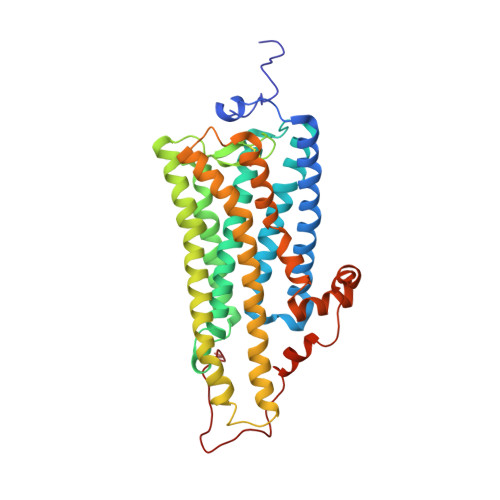
Crystal structure of squid rhodopsin with intracellularly extended cytoplasmic region

Shimamura, T.Hiraki, K.Takahashi, N.Hori, T.Ago, H.Masuda, K.Takio, K.Ishiguro, M.Miyano, M.

(2008) J Biol Chem 283: 17753-17756

  • DOI: https://doi.org/10.1074/jbc.C800040200
  • Primary Citation of Related Structures:  
    2ZIY

  • PubMed Abstract: 
  • G-protein-coupled receptors play a key step in cellular signal transduction cascades by transducing various extracellular signals via G-proteins. Rhodopsin is a prototypical G-protein-coupled receptor involved in the retinal visual signaling cascade. We determined the structure of squid rhodopsin at 3 ...

    G-protein-coupled receptors play a key step in cellular signal transduction cascades by transducing various extracellular signals via G-proteins. Rhodopsin is a prototypical G-protein-coupled receptor involved in the retinal visual signaling cascade. We determined the structure of squid rhodopsin at 3.7A resolution, which transduces signals through the G(q) protein to the phosphoinositol cascade. The structure showed seven transmembrane helices and an amphipathic helix H8 has similar geometry to structures from bovine rhodopsin, coupling to G(t), and human beta(2)-adrenergic receptor, coupling to G(s). Notably, squid rhodopsin contains a well structured cytoplasmic region involved in the interaction with G-proteins, and this region is flexible or disordered in bovine rhodopsin and human beta(2)-adrenergic receptor. The transmembrane helices 5 and 6 are longer and extrude into the cytoplasm. The distal C-terminal tail contains a short hydrophilic alpha-helix CH after the palmitoylated cysteine residues. The residues in the distal C-terminal tail interact with the neighboring residues in the second cytoplasmic loop, the extruded transmembrane helices 5 and 6, and the short helix H8. Additionally, the Tyr-111, Asn-87, and Asn-185 residues are located within hydrogen-bonding distances from the nitrogen atom of the Schiff base.


    Organizational Affiliation

    RIKEN SPring-8 Center, Harima Institute, Kouto, Sayo, Hyogo, Japan.


Macromolecules
Find similar proteins by:  (by identity cutoff)  |  3D Structure
Entity ID: 1
MoleculeChainsSequence LengthOrganismDetailsImage
RhodopsinA372Todarodes pacificusMutation(s): 0 
Gene Names: RHO
Membrane Entity: Yes 
UniProt
Find proteins for P31356 (Todarodes pacificus)
Explore P31356 
Go to UniProtKB:  P31356
Entity Groups  
Sequence Clusters30% Identity50% Identity70% Identity90% Identity95% Identity100% Identity
UniProt GroupP31356
Protein Feature View
Expand
  • Reference Sequence
Small Molecules
Ligands 2 Unique
IDChainsName / Formula / InChI Key2D Diagram3D Interactions
RET
Query on RET

Download Ideal Coordinates CCD File 
B [auth A]RETINAL
C20 H28 O
NCYCYZXNIZJOKI-OVSJKPMPSA-N
 Ligand Interaction
PLM
Query on PLM

Download Ideal Coordinates CCD File 
C [auth A],
D [auth A]
PALMITIC ACID
C16 H32 O2
IPCSVZSSVZVIGE-UHFFFAOYSA-N
 Ligand Interaction
Experimental Data & Validation

Experimental Data

  • Method: X-RAY DIFFRACTION
  • Resolution: 3.70 Å
  • R-Value Free: 0.330 
  • R-Value Work: 0.302 
  • Space Group: C 2 2 21
Unit Cell:
Length ( Å )Angle ( ˚ )
a = 84.319α = 90
b = 108.746β = 90
c = 142.165γ = 90
Software Package:
Software NamePurpose
MOLREPphasing
CNSrefinement
HKL-2000data reduction
HKL-2000data scaling

Structure Validation

View Full Validation Report


Ligand Structure Quality Assessment 


Entry History 

Deposition Data

Revision History  (Full details and data files)

  • Version 1.0: 2008-05-06
    Type: Initial release
  • Version 1.1: 2011-07-13
    Changes: Version format compliance