2YQK | pdb_00002yqk

Solution structure of the SANT domain in Arginine-glutamic acid dipeptide (RE) repeats


Experimental Data Snapshot

  • Method: SOLUTION NMR
  • Conformers Calculated: 100 
  • Conformers Submitted: 20 
  • Selection Criteria: structures with the least restraint violations,target function 

wwPDB Validation 3D Report Full Report

Validation slider image for 2YQK

This is version 1.3 of the entry. See complete history

Literature

Solution structure of the SANT domain in Arginine-glutamic acid dipeptide (RE) repeats

Kadirvel, S.He, F.Muto, Y.Inoue, M.Kigawa, T.Shirouzu, M.Tarada, T.Yokoyama, S.

To be published.

Macromolecule Content 

  • Total Structure Weight: 7.3 kDa 
  • Atom Count: 515 
  • Modeled Residue Count: 63 
  • Deposited Residue Count: 63 
  • Unique protein chains: 1

Macromolecules

Find similar proteins by:|  3D Structure
Entity ID: 1
MoleculeChains  Sequence LengthOrganismDetailsImage
Arginine-glutamic acid dipeptide repeats protein63Homo sapiensMutation(s): 0 
Gene Names: RERE
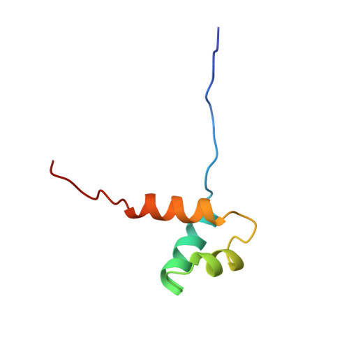
UniProt & NIH Common Fund Data Resources
Find proteins for Q9P2R6 (Homo sapiens)
Explore Q9P2R6 
Go to UniProtKB:  Q9P2R6
PHAROS:  Q9P2R6
GTEx:  ENSG00000142599 
Entity Groups
Sequence Clusters30% Identity50% Identity70% Identity90% Identity95% Identity100% Identity
UniProt GroupQ9P2R6
Sequence Annotations
Expand
Reference Sequence

Experimental Data & Validation

Experimental Data

  • Method: SOLUTION NMR
  • Conformers Calculated: 100 
  • Conformers Submitted: 20 
  • Selection Criteria: structures with the least restraint violations,target function 

Structure Validation

View Full Validation Report


Entry History 

Revision History  (Full details and data files)

  • Version 1.0: 2007-10-02
    Type: Initial release
  • Version 1.1: 2011-07-13
    Changes: Version format compliance
  • Version 1.2: 2022-03-16
    Changes: Data collection, Database references, Derived calculations
  • Version 1.3: 2024-05-29
    Changes: Data collection