Evidence for a Two-Metal-Ion Mechanism in the Cytidyltransferase Kdsb, an Enzyme Involved in Lipopolysaccharide Biosynthesis.
Schmidt, H., Mesters, J.R., Wu, J., Woodard, R.W., Hilgenfeld, R., Mamat, U.(2011) PLoS One 6: 23231
- PubMed: 21826242 Search on PubMedSearch on PubMed Central
- DOI: https://doi.org/10.1371/journal.pone.0023231
- Primary Citation Related Structures:
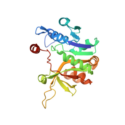
2Y6P - PubMed Abstract:
Lipopolysaccharide (LPS) is located on the surface of Gram-negative bacteria and is responsible for maintaining outer membrane stability, which is a prerequisite for cell survival. Furthermore, it represents an important barrier against hostile environmental factors such as antimicrobial peptides and the complement cascade during Gram-negative infections. The sugar 3-deoxy-D-manno-oct-2-ulosonic acid (Kdo) is an integral part of LPS and plays a key role in LPS functionality. Prior to its incorporation into the LPS molecule, Kdo has to be activated by the CMP-Kdo synthetase (CKS). Based on the presence of a single Mg²⁺ ion in the active site, detailed models of the reaction mechanism of CKS have been developed previously. Recently, a two-metal-ion hypothesis suggested the involvement of two Mg²⁺ ions in Kdo activation. To further investigate the mechanistic aspects of Kdo activation, we kinetically characterized the CKS from the hyperthermophilic organism Aquifex aeolicus. In addition, we determined the crystal structure of this enzyme at a resolution of 2.10 Å and provide evidence that two Mg²⁺ ions are part of the active site of the enzyme.
- Institute of Biochemistry, Center for Structural and Cell Biology in Medicine, University of Lübeck, Lübeck, Germany.
Organizational Affiliation: