2Y5T | pdb_00002y5t

Crystal structure of the pathogenic autoantibody CIIC1 in complex with the triple-helical C1 peptide


Experimental Data Snapshot

  • Method: X-RAY DIFFRACTION
  • Resolution: 2.20 Å
  • R-Value Free: 
    0.245 (Depositor), 0.263 (DCC) 
  • R-Value Work: 
    0.188 (Depositor), 0.209 (DCC) 
  • R-Value Observed: 
    0.191 (Depositor) 

Starting Model: experimental
View more details

wwPDB Validation 3D Report Full Report

Validation slider image for 2Y5T

This is version 1.2 of the entry. See complete history

Literature

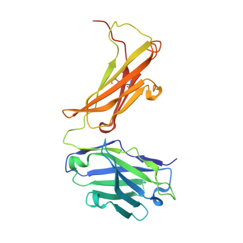
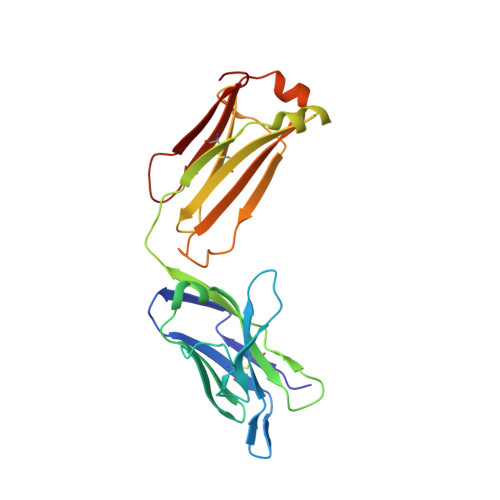
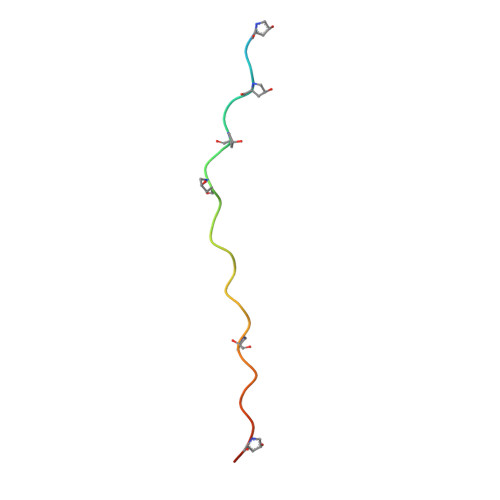
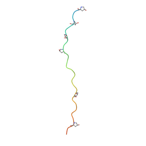
Crystal Structure of an Arthritogenic Anticollagen Immune Complex.

Dobritzsch, D.Lindh, I.Uysal, H.Nandakumar, K.S.Burkhardt, H.Schneider, G.Holmdahl, R.

(2011) Arthritis Rheum 63: 3740

  • DOI: https://doi.org/10.1002/art.30611
  • Primary Citation Related Structures: 
    2Y5T

  • PubMed Abstract: 

    In rheumatoid arthritis, joint inflammation and cartilage destruction are mediated by autoantibodies directed to various self antigens. Type II collagen (CII)-specific antibodies are likely to play a role in this process and have been shown to induce experimental arthritis in susceptible animals. The purpose of this study was to reveal how arthritogenic autoantibodies recognize native CII in its triple-helical conformation. Site-directed mutagenesis and crystallographic studies were performed to reveal crucial contact points between the CII antibody and the triple-helical CII peptide. The crystal structure of a pathogenic autoantibody bound to a major triple-helical epitope present on CII was determined, allowing a first and detailed description of the interactions within an arthritogenic complex that is frequently occurring in both mice and humans with autoimmune arthritis. The crystal structure emphasizes the role of arginine residues located in a commonly recognized motif on CII and reveals that germline-encoded elements are involved in the interaction with the epitope. The crystal structure of an arthritogenic antibody binding a triple-helical epitope on CII indicates a crucial role of germline-encoded and arginine residues as the target structures.


  • Organizational Affiliation
    • Karolinska Institute, Stockholm, Sweden.

Macromolecule Content 

  • Total Structure Weight: 58.87 kDa 
  • Atom Count: 4,229 
  • Modeled Residue Count: 517 
  • Deposited Residue Count: 556 
  • Unique protein chains: 4

Macromolecules

Find similar proteins by:|  3D Structure
Entity ID: 1
MoleculeChains  Sequence LengthOrganismDetailsImage
CIIC1 FAB FRAGMENT HEAVY CHAIN232Mus musculusMutation(s): 0 
Find similar proteins by:|  3D Structure
Entity ID: 2
MoleculeChains  Sequence LengthOrganismDetailsImage
CIIC1 FAB FRAGMENT LIGHT CHAIN218Mus musculusMutation(s): 0 
Find similar proteins by:|  3D Structure
Entity ID: 3
MoleculeChains  Sequence LengthOrganismDetailsImage
C1C [auth E],
E [auth G]
34Mus musculusMutation(s): 0 
Find similar proteins by:|  3D Structure
Entity ID: 4
MoleculeChains  Sequence LengthOrganismDetailsImage
C1D [auth F]38Mus musculusMutation(s): 0 

Small Molecules

Experimental Data & Validation

Experimental Data

  • Method: X-RAY DIFFRACTION
  • Resolution: 2.20 Å
  • R-Value Free:  0.245 (Depositor), 0.263 (DCC) 
  • R-Value Work:  0.188 (Depositor), 0.209 (DCC) 
  • R-Value Observed: 0.191 (Depositor) 
Space Group: P 31 2 1
Unit Cell:
Length ( Å )Angle ( ˚ )
a = 81.05α = 90
b = 81.05β = 90
c = 168.45γ = 120
Software Package:
Software NamePurpose
REFMACrefinement
iMOSFLMdata reduction
SCALAdata scaling
PHASERphasing

Structure Validation

View Full Validation Report


Entry History 

Deposition Data

Revision History  (Full details and data files)

  • Version 1.0: 2011-12-14
    Type: Initial release
  • Version 1.1: 2017-08-23
    Changes: Data collection
  • Version 1.2: 2023-12-20
    Changes: Data collection, Database references, Derived calculations, Other, Refinement description