2Y2Q | pdb_00002y2q

PENICILLIN-BINDING PROTEIN 1B (PBP-1B) IN COMPLEX WITH AN ALKYL BORONATE (Z06)


Experimental Data Snapshot

  • Method: X-RAY DIFFRACTION
  • Resolution: 2.15 Å
  • R-Value Free: 
    0.262 (Depositor), 0.262 (DCC) 
  • R-Value Work: 
    0.203 (Depositor), 0.204 (DCC) 
  • R-Value Observed: 
    0.206 (Depositor) 

Starting Model: experimental
View more details

wwPDB Validation 3D Report Full Report

Validation slider image for 2Y2Q

Ligand Structure Quality Assessment 


This is version 1.3 of the entry. See complete history

Literature

Structure-Guided Design of Cell Wall Biosynthesis Inhibitors that Overcome Beta-Lactam Resistance in Staphylococcus Aureus (Mrsa).

Contreras-Martel, C.Amoroso, A.Woon, E.C.Zervosen, A.Inglis, S.Martins, A.Verlaine, O.Rydzik, A.Job, V.Luxen, A.Joris, B.Schofield, C.J.Dessen, A.

(2011) ACS Chem Biol 6: 943

  • DOI: https://doi.org/10.1021/cb2001846
  • Primary Citation Related Structures: 
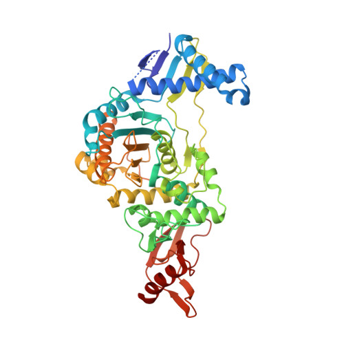
    2Y2G, 2Y2H, 2Y2I, 2Y2J, 2Y2K, 2Y2L, 2Y2M, 2Y2N, 2Y2O, 2Y2P, 2Y2Q

  • PubMed Abstract: 

    β-Lactam antibiotics have long been a treatment of choice for bacterial infections since they bind irreversibly to Penicillin-Binding Proteins (PBPs), enzymes that are vital for cell wall biosynthesis. Many pathogens express drug-insensitive PBPs rendering β-lactams ineffective, revealing a need for new types of PBP inhibitors active against resistant strains. We have identified alkyl boronic acids that are active against pathogens including methicillin-resistant S. aureus (MRSA). The crystal structures of PBP1b complexed to 11 different alkyl boronates demonstrate that in vivo efficacy correlates with the mode of inhibitor side chain binding. Staphylococcal membrane analyses reveal that the most potent alkyl boronate targets PBP1, an autolysis system regulator, and PBP2a, a low β-lactam affinity enzyme. This work demonstrates the potential of boronate-based PBP inhibitors for circumventing β-lactam resistance and opens avenues for the development of novel antibiotics that target Gram-positive pathogens.


  • Organizational Affiliation
    • Bacterial Pathogenesis Group, Institut de Biologie Structurale, Université Grenoble I, France.

Macromolecule Content 

  • Total Structure Weight: 109.88 kDa 
  • Atom Count: 7,870 
  • Modeled Residue Count: 943 
  • Deposited Residue Count: 988 
  • Unique protein chains: 1

Macromolecules

Find similar proteins by:|  3D Structure
Entity ID: 1
MoleculeChains  Sequence LengthOrganismDetailsImage
PENICILLIN-BINDING PROTEIN 1B
A, B
494Streptococcus pneumoniae R6Mutation(s): 3 
EC: 2.4.1.129 (PDB Primary Data), 2.3.2 (PDB Primary Data)
UniProt
Find proteins for Q7CRA4 (Streptococcus pneumoniae (strain ATCC BAA-255 / R6))
Explore Q7CRA4 
Go to UniProtKB:  Q7CRA4
Entity Groups
Sequence Clusters30% Identity50% Identity70% Identity90% Identity95% Identity100% Identity
UniProt GroupQ7CRA4
Sequence Annotations
Expand
Reference Sequence

Small Molecules

Ligands 5 Unique
IDChains Name / Formula / InChI Key2D Diagram3D Interactions
Z06

Query on Z06


Download:Ideal Coordinates CCD File
C [auth A],
T [auth B]
(3-acetamido-5-carboxy-phenyl)-trihydroxy-boron
C9 H11 B N O6
ZONDIXDEBAKRPT-UHFFFAOYSA-N
SO4

Query on SO4


Download:Ideal Coordinates CCD File
D [auth A],
U [auth B]
SULFATE ION
O4 S
QAOWNCQODCNURD-UHFFFAOYSA-L
EDO

Query on EDO


Download:Ideal Coordinates CCD File
R [auth A]1,2-ETHANEDIOL
C2 H6 O2
LYCAIKOWRPUZTN-UHFFFAOYSA-N
CL

Query on CL


Download:Ideal Coordinates CCD File
AA [auth B]
BA [auth B]
CA [auth B]
DA [auth B]
E [auth A]
AA [auth B],
BA [auth B],
CA [auth B],
DA [auth B],
E [auth A],
EA [auth B],
F [auth A],
FA [auth B],
G [auth A],
GA [auth B],
H [auth A],
I [auth A],
J [auth A],
K [auth A],
L [auth A],
M [auth A],
N [auth A],
O [auth A],
P [auth A],
S [auth A],
V [auth B],
W [auth B],
X [auth B],
Y [auth B],
Z [auth B]
CHLORIDE ION
Cl
VEXZGXHMUGYJMC-UHFFFAOYSA-M
NA

Query on NA


Download:Ideal Coordinates CCD File
HA [auth B],
IA [auth B],
Q [auth A]
SODIUM ION
Na
FKNQFGJONOIPTF-UHFFFAOYSA-N

Experimental Data & Validation

Experimental Data

  • Method: X-RAY DIFFRACTION
  • Resolution: 2.15 Å
  • R-Value Free:  0.262 (Depositor), 0.262 (DCC) 
  • R-Value Work:  0.203 (Depositor), 0.204 (DCC) 
  • R-Value Observed: 0.206 (Depositor) 
Space Group: P 21 21 21
Unit Cell:
Length ( Å )Angle ( ˚ )
a = 96.152α = 90
b = 101.806β = 90
c = 145.37γ = 90
Software Package:
Software NamePurpose
REFMACrefinement
XDSdata reduction
XSCALEdata scaling
PHASERphasing

Structure Validation

View Full Validation Report


Ligand Structure Quality Assessment 


Entry History 

Revision History  (Full details and data files)

  • Version 1.0: 2011-08-03
    Type: Initial release
  • Version 1.1: 2011-09-28
    Changes: Database references
  • Version 1.2: 2023-12-20
    Changes: Data collection, Database references, Derived calculations, Other, Refinement description
  • Version 1.3: 2024-10-23
    Changes: Structure summary