2XSA

OgOGA apostructure


Experimental Data Snapshot

  • Method: X-RAY DIFFRACTION
  • Resolution: 2.00 Å
  • R-Value Free: 0.223 
  • R-Value Work: 0.186 
  • R-Value Observed: 0.187 

wwPDB Validation   3D Report Full Report


This is version 1.2 of the entry. See complete history


Literature

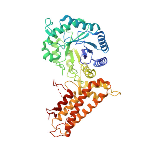
Human Oga Binds Substrates in a Conserved Peptide Recognition Groove.

Schimpl, M.Schuttelkopf, A.W.Borodkin, V.S.Van Aalten, D.M.F.

(2010) Biochem J 432: 1

  • DOI: https://doi.org/10.1042/BJ20101338
  • Primary Citation of Related Structures:  
    2XSA, 2XSB

  • PubMed Abstract: 

    Modification of cellular proteins with O-GlcNAc (O-linked N-acetylglucosamine) competes with protein phosphorylation and regulates a plethora of cellular processes. O-GlcNAcylation is orchestrated by two opposing enzymes, O-GlcNAc transferase and OGA (O-GlcNAcase or β-N-acetylglucosaminidase), which recognize their target proteins via as yet unidentified mechanisms. In the present study, we uncovered the first insights into the mechanism of substrate recognition by human OGA. The structure of a novel bacterial OGA orthologue reveals a putative substrate-binding groove, conserved in metazoan OGAs. Guided by this structure, conserved amino acids lining this groove in human OGA were mutated and the activity on three different substrate proteins [TAB1 (transforming growth factor-β-activated protein kinase 1-binding protein 1), FoxO1 (forkhead box O1) and CREB (cAMP-response-element-binding protein)] was tested in an in vitro deglycosylation assay. The results provide the first evidence that human OGA may possess a substrate-recognition mechanism that involves interactions with O-GlcNAcylated proteins beyond the GlcNAc-binding site, with possible implications for differential regulation of cycling of O-GlcNAc on different proteins.


  • Organizational Affiliation

    University of Dundee, Scotland, UK.


Macromolecules
Find similar proteins by:  (by identity cutoff)  |  3D Structure
Entity ID: 1
MoleculeChains Sequence LengthOrganismDetailsImage
HYALURONOGLUCOSAMINIDASE447Oceanicola granulosus HTCC2516Mutation(s): 0 
EC: 3.2.1.52
UniProt
Find proteins for Q2CEE3 (Oceanicola granulosus (strain ATCC BAA-861 / DSM 15982 / KCTC 12143 / HTCC2516))
Explore Q2CEE3 
Go to UniProtKB:  Q2CEE3
Entity Groups  
Sequence Clusters30% Identity50% Identity70% Identity90% Identity95% Identity100% Identity
UniProt GroupQ2CEE3
Sequence Annotations
Expand
  • Reference Sequence
Small Molecules
Ligands 1 Unique
IDChains Name / Formula / InChI Key2D Diagram3D Interactions
CL
Query on CL

Download Ideal Coordinates CCD File 
B [auth A]CHLORIDE ION
Cl
VEXZGXHMUGYJMC-UHFFFAOYSA-M
Experimental Data & Validation

Experimental Data

  • Method: X-RAY DIFFRACTION
  • Resolution: 2.00 Å
  • R-Value Free: 0.223 
  • R-Value Work: 0.186 
  • R-Value Observed: 0.187 
  • Space Group: C 1 2 1
Unit Cell:
Length ( Å )Angle ( ˚ )
a = 84.396α = 90
b = 95.349β = 104.01
c = 60.528γ = 90
Software Package:
Software NamePurpose
REFMACrefinement
DENZOdata reduction
SCALEPACKdata scaling

Structure Validation

View Full Validation Report


Entry History 

Deposition Data

Revision History  (Full details and data files)

  • Version 1.0: 2011-03-23
    Type: Initial release
  • Version 1.1: 2011-05-08
    Changes: Version format compliance
  • Version 1.2: 2011-07-13
    Changes: Version format compliance