2XQZ | pdb_00002xqz

Neutron structure of the perdeuterated Toho-1 R274N R276N double mutant beta-lactamase


Experimental Data Snapshot

  • Method: NEUTRON DIFFRACTION
  • Resolution: 2.10 Å
  • R-Value Free: 
    0.259 (Depositor) 
  • R-Value Work: 
    0.225 (Depositor) 
  • R-Value Observed: 
    0.228 (Depositor) 

wwPDB Validation 3D Report Full Report

Validation slider image for 2XQZ

This is version 2.2 of the entry. See complete history

Literature

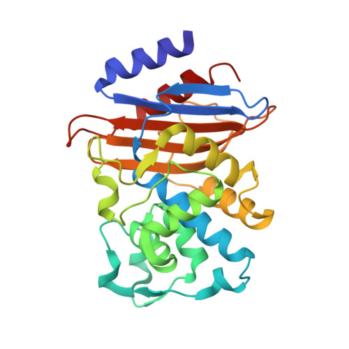
The Active Site Protonation States of Perdeuterated Toho-1 Beta-Lactamase Determined by Neutron Diffraction Support a Role for Glu166 as the General Base in Acylation.

Tomanicek, S.J.Wang, K.K.Weiss, K.L.Blakeley, M.P.Cooper, J.Chen, Y.Coates, L.

(2011) FEBS Lett 585: 364

  • DOI: https://doi.org/10.1016/j.febslet.2010.12.017
  • Primary Citation Related Structures: 
    2XQZ, 2XR0

  • PubMed Abstract: 

    Room temperature neutron diffraction data of the fully perdeuterated Toho-1 R274N/R276N double mutant β-lactamase in the apo form were used to visualize deuterium atoms within the active site of the enzyme. This perdeuterated neutron structure of the Toho-1 R274N/R276N reveals the clearest picture yet of the ground-state active site protonation states and the complete hydrogen-bonding network in a β-lactamase enzyme. The ground-state active site protonation states detailed in this neutron diffraction study are consistent with previous high-resolution X-ray studies that support the role of Glu166 as the general base during the acylation reaction in the class A β-lactamase reaction pathway.


  • Organizational Affiliation
    • Oak Ridge National Laboratory, Neutron Scattering Science Division, Oak Ridge, TN 37831, USA.

Macromolecule Content 

  • Total Structure Weight: 27.99 kDa 
  • Atom Count: 2,169 
  • Modeled Residue Count: 260 
  • Deposited Residue Count: 260 
  • Unique protein chains: 1

Macromolecules

Find similar proteins by:|  3D Structure
Entity ID: 1
MoleculeChains  Sequence LengthOrganismDetailsImage
BETA-LACTAMSE TOHO-1260Escherichia coliMutation(s): 2 
EC: 3.5.2.6
UniProt
Find proteins for Q47066 (Escherichia coli)
Explore Q47066 
Go to UniProtKB:  Q47066
Entity Groups
Sequence Clusters30% Identity50% Identity70% Identity90% Identity95% Identity100% Identity
UniProt GroupQ47066
Sequence Annotations
Expand
Reference Sequence

Experimental Data & Validation

Experimental Data

  • Method: NEUTRON DIFFRACTION
  • Resolution: 2.10 Å
  • R-Value Free:  0.259 (Depositor) 
  • R-Value Work:  0.225 (Depositor) 
  • R-Value Observed: 0.228 (Depositor) 
Space Group: P 32 2 1
Unit Cell:
Length ( Å )Angle ( ˚ )
a = 72.956α = 90
b = 72.956β = 90
c = 98.526γ = 120
Software Package:
Software NamePurpose
PHENIXrefinement
LAUEGENdata reduction
LAUEGENdata scaling

Structure Validation

View Full Validation Report


Entry History 

Deposition Data

Revision History  (Full details and data files)

  • Version 1.0: 2010-12-22
    Type: Initial release
  • Version 2.0: 2017-12-27
    Changes: Advisory, Atomic model, Derived calculations, Other
  • Version 2.1: 2018-01-31
    Changes: Data collection
  • Version 2.2: 2024-05-08
    Changes: Data collection, Database references