Structure of Cytochrome P450 Pimd Suggests Epoxidation of the Polyene Macrolide Pimaricin Occurs Via a Hydroperoxoferric Intermediate.
Kells, P.M., Ouellet, H., Santos-Aberturas, J., Aparicio, J.F., Podust, L.M.(2010) Chem Biol 17: 841
- PubMed: 20797613 Search on PubMedSearch on PubMed Central
- DOI: https://doi.org/10.1016/j.chembiol.2010.05.026
- Primary Citation Related Structures:
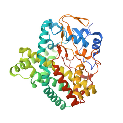
2X9P, 2XBK - PubMed Abstract:
We present the X-ray structure of PimD, both substrate-free and in complex with 4,5-desepoxypimaricin. PimD is a cytochrome P450 monooxygenase with native epoxidase activity that is critical in the biosynthesis of the polyene macrolide antibiotic pimaricin. Intervention in this secondary metabolic pathway could advance the development of drugs with improved pharmacologic properties. Epoxidation by P450 typically includes formation of a charge-transfer complex between an oxoferryl pi-cation radical species (Compound I) and the olefin pi-bond as the initial intermediate. Catalytic and structural evidence presented here suggest that epoxidation of 4,5-desepoxypimaricin proceeds via a hydroperoxoferric intermediate (Compound 0). The oxygen atom of Compound 0 distal to the heme iron may insert into the double bond of the substrate to make an epoxide ring. Stereoelectronic features of the putative transition state suggest substrate-assisted proton delivery.
- Department of Pharmaceutical Chemistry, University of California, San Francisco, CA 94158, USA.
Organizational Affiliation: