Structural and Mechanistic Insight Into the Ferredoxin-Mediated Two-Electron Reduction of Bilins.
Busch, A.W.U., Reijerse, E.J., Lubitz, W., Frankenberg-Dinkel, N., Hofmann, E.(2011) Biochem J 439: 257
- PubMed: 21729003 Search on PubMed
- DOI: https://doi.org/10.1042/BJ20110814
- Primary Citation Related Structures:
2X9O - PubMed Abstract:
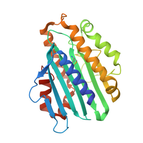
PEB (phycoerythrobilin) is one of the major open-chain tetrapyrrole molecules found in cyanobacterial light-harvesting phycobiliproteins. In these organisms, two enzymes of the ferredoxin-dependent bilin reductase family work in tandem to reduce BV (biliverdin IXα) to PEB. In contrast, a single cyanophage-encoded enzyme of the same family has been identified to catalyse the identical reaction. Using UV-visible and EPR spectroscopy we investigated the two individual cyanobacterial enzymes PebA [15,16-DHBV (dihydrobiliverdin):ferredoxin oxidoreductase] and PebB (PEB:ferredoxin oxidoreductase) showing that the two subsequent reactions catalysed by the phage enzyme PebS (PEB synthase) are clearly dissected in the cyanobacterial versions. Although a highly conserved aspartate residue is critical for both reductions, a second conserved aspartate residue is only involved in the A-ring reduction of the tetrapyrrole in PebB and PebS. The crystal structure of PebA from Synechococcus sp. WH8020 in complex with its substrate BV at a 1.55 Å (1 Å=0.1 nm) resolution revealed further insight into the understanding of enzyme evolution and function. Based on the structure it becomes obvious that in addition to the importance of certain catalytic residues, the shape of the active site and consequently the binding of the substrate highly determines the catalytic properties.
- Physiology of Microorganisms, Faculty of Biology and Biotechnology, Ruhr-University Bochum, 44780 Bochum, Germany.
Organizational Affiliation: