2WYF

Crystal structure of PA-IL lectin complexed with aGal12bGal-O-Met at 2.4 A resolution


Experimental Data Snapshot

  • Method: X-RAY DIFFRACTION
  • Resolution: 2.40 Å
  • R-Value Free: 0.259 
  • R-Value Work: 0.166 
  • R-Value Observed: 0.170 

wwPDB Validation   3D Report Full Report


This is version 2.1 of the entry. See complete history


Literature

Role of Water Molecules in Structure and Energetics of Pseudomonas Aeruginosa Lectin I Interacting with Disaccharides.

Nurisso, A.Blanchard, B.Audfray, A.Rydner, L.Oscarson, S.Varrot, A.Imberty, A.

(2010) J Biol Chem 285: 20316

  • DOI: https://doi.org/10.1074/jbc.M110.108340
  • Primary Citation of Related Structures:  
    2WYF

  • PubMed Abstract: 

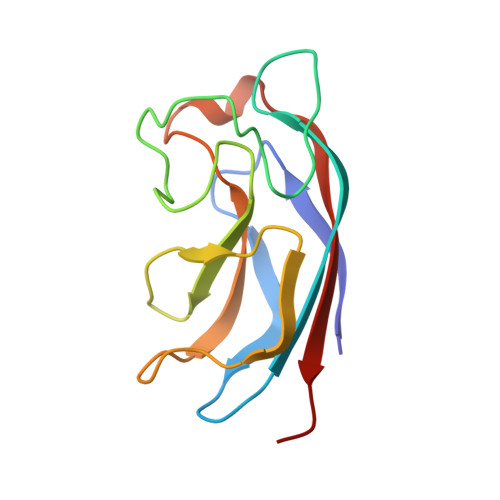
    Calcium-dependent lectin I from Pseudomonas aeruginosa (PA-IL) binds specifically to oligosaccharides presenting an alpha-galactose residue at their nonreducing end, such as the disaccharides alphaGal1-2betaGalOMe, alphaGal1-3betaGalOMe, and alphaGal1-4betaGalOMe. This provides a unique model for studying the effect of the glycosidic linkage of the ligands on structure and thermodynamics of the complexes by means of experimental and theoretical tools. The structural features of PA-IL in complex with the three disaccharides were established by docking and molecular dynamics simulations and compared with those observed in available crystal structures, including PA-IL.alphaGal1-2betaGalOMe complex, which was solved at 2.4 A resolution and reported herein. The role of a structural bridge water molecule in the binding site of PA-IL was also elucidated through molecular dynamics simulations and free energy calculations. This water molecule establishes three very stable hydrogen bonds with O6 of nonreducing galactose, oxygen from Pro-51 main chain, and nitrogen from Gln-53 main chain of the lectin binding site. Binding free energies for PA-IL in complex with the three disaccharides were investigated, and the results were compared with the experimental data determined by titration microcalorimetry. When the bridge water molecule was included in the free energy calculations, the simulations predicted the correct binding affinity trends with the 1-2-linked disaccharide presenting three times stronger affinity ligand than the other two. These results highlight the role of the water molecule in the binding site of PA-IL and indicate that it should be taken into account when designing glycoderivatives active against P. aeruginosa adhesion.


  • Organizational Affiliation

    Centre de Rechèrche sur les Macromolécules Végétales-CNRS (affiliated with Université Joseph Fourier and Institut de Chimie Moléculaire de Grenoble), BP 53, 38041 Grenoble Cedex 9, France.


Macromolecules
Find similar proteins by:  (by identity cutoff)  |  3D Structure
Entity ID: 1
MoleculeChains Sequence LengthOrganismDetailsImage
PA-I GALACTOPHILIC LECTIN
A, B, C, D, E
A, B, C, D, E, F, G, H
121Pseudomonas aeruginosa PAO1Mutation(s): 0 
UniProt
Find proteins for Q05097 (Pseudomonas aeruginosa (strain ATCC 15692 / DSM 22644 / CIP 104116 / JCM 14847 / LMG 12228 / 1C / PRS 101 / PAO1))
Explore Q05097 
Go to UniProtKB:  Q05097
Entity Groups  
Sequence Clusters30% Identity50% Identity70% Identity90% Identity95% Identity100% Identity
UniProt GroupQ05097
Sequence Annotations
Expand
  • Reference Sequence
Oligosaccharides

Help

Entity ID: 2
MoleculeChains Length2D Diagram Glycosylation3D Interactions
alpha-D-galactopyranose-(1-2)-methyl beta-D-galactopyranoside
I, J, K, L, M
I, J, K, L, M, N
2N/A
Glycosylation Resources
GlyTouCan:  G50103OC
GlyCosmos:  G50103OC
Small Molecules
Ligands 2 Unique
IDChains Name / Formula / InChI Key2D Diagram3D Interactions
GLA
Query on GLA

Download Ideal Coordinates CCD File 
Q [auth B],
U [auth E]
alpha-D-galactopyranose
C6 H12 O6
WQZGKKKJIJFFOK-PHYPRBDBSA-N
CA
Query on CA

Download Ideal Coordinates CCD File 
O [auth A]
P [auth B]
R [auth C]
S [auth D]
T [auth E]
O [auth A],
P [auth B],
R [auth C],
S [auth D],
T [auth E],
V [auth F],
W [auth G],
X [auth H]
CALCIUM ION
Ca
BHPQYMZQTOCNFJ-UHFFFAOYSA-N
Binding Affinity Annotations 
IDSourceBinding Affinity
MBG BindingDB:  2WYF Kd: 7.00e+4 (nM) from 1 assay(s)
Experimental Data & Validation

Experimental Data

  • Method: X-RAY DIFFRACTION
  • Resolution: 2.40 Å
  • R-Value Free: 0.259 
  • R-Value Work: 0.166 
  • R-Value Observed: 0.170 
  • Space Group: P 1 21 1
Unit Cell:
Length ( Å )Angle ( ˚ )
a = 49.885α = 90
b = 99.788β = 100.77
c = 91.318γ = 90
Software Package:
Software NamePurpose
REFMACrefinement
MOSFLMdata reduction
SCALAdata scaling
PHASERphasing

Structure Validation

View Full Validation Report


Entry History 

Deposition Data

Revision History  (Full details and data files)

  • Version 1.0: 2010-04-21
    Type: Initial release
  • Version 1.1: 2011-08-03
    Changes: Database references, Derived calculations, Non-polymer description, Source and taxonomy, Version format compliance
  • Version 2.0: 2020-07-29
    Type: Remediation
    Reason: Carbohydrate remediation
    Changes: Atomic model, Data collection, Derived calculations, Other, Structure summary
  • Version 2.1: 2023-12-20
    Changes: Data collection, Database references, Refinement description, Structure summary