2WXD | pdb_00002wxd

A MICROMOLAR O-SULFATED THIOHYDROXIMATE INHIBITOR BOUND TO PLANT MYROSINASE


Experimental Data Snapshot

  • Method: X-RAY DIFFRACTION
  • Resolution: 1.60 Å
  • R-Value Free: 
    0.164 (Depositor), 0.174 (DCC) 
  • R-Value Work: 
    0.143 (Depositor), 0.153 (DCC) 
  • R-Value Observed: 
    0.144 (Depositor) 

Starting Model: experimental
View more details

wwPDB Validation 3D Report Full Report

Validation slider image for 2WXD

Ligand Structure Quality Assessment 


This is version 2.2 of the entry. See complete history

Literature

A Micromolar O-Sulfated Thiohydroximate Inhibitor Bound to Plant Myrosinase

Besle, A.Brazzolotto, X.Tatibouet, A.Cerniauskaite, D.Gallienne, E.Rollin, P.Burmeister, W.P.

(2010) Acta Crystallogr Sect F Struct Biol Cryst Commun 66: 152

  • DOI: https://doi.org/10.1107/S1744309109052865
  • Primary Citation Related Structures: 
    2WXD

  • PubMed Abstract: 

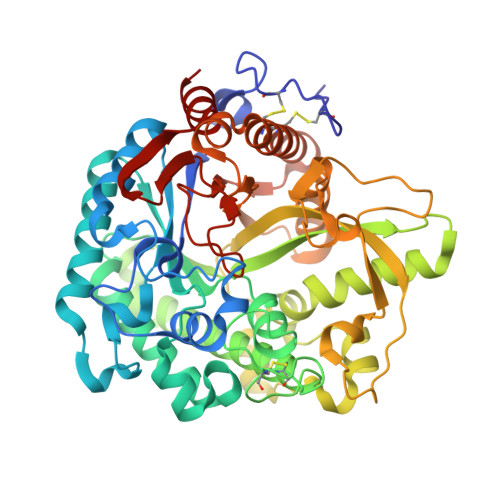
    The 1.6 A resolution structure of the micromolar competitive inhibitor S-(N,N-dimethylaminoethyl) phenylacetothiohydroximate-O-sulfate bound to Sinapis alba myrosinase, a plant thioglucosidase, is reported. Myrosinase and its substrates, the glucosinolates, are part of the plant's defence system. The sulfate group and the phenyl group of the inhibitor bind to the aglycon-binding site of the enzyme, whereas the N,N-dimethyl group binds to the glucose-binding site and explains the large improvement in binding affinity compared with previous compounds. The structure suggests ways to increase the potency and specificity of the compound by improving the interactions with the hydrophobic pocket of the aglycon-binding site.


  • Organizational Affiliation
    • UJF-EMBL-CNRS UMI 3265, Unit of Virus Host Cell Interactions, BP 181, France.

Macromolecule Content 

  • Total Structure Weight: 63.14 kDa 
  • Atom Count: 5,238 
  • Modeled Residue Count: 499 
  • Deposited Residue Count: 501 
  • Unique protein chains: 1

Macromolecules

Find similar proteins by:|  3D Structure
Entity ID: 1
MoleculeChains  Sequence LengthOrganismDetailsImage
MYROSINASEA [auth M]501Sinapis albaMutation(s): 0 
EC: 3.2.3.1 (PDB Primary Data), 3.2.1.147 (UniProt)
UniProt
Find proteins for P29736 (Sinapis alba)
Explore P29736 
Go to UniProtKB:  P29736
Entity Groups
Sequence Clusters30% Identity50% Identity70% Identity90% Identity95% Identity100% Identity
UniProt GroupP29736
Glycosylation
Glycosylation Sites: 10
Sequence Annotations
Expand
Reference Sequence

Oligosaccharides

Help  
Entity ID: 2
MoleculeChains Length2D Diagram GlycosylationD Interactions
2-acetamido-2-deoxy-beta-D-glucopyranose-(1-4)-2-acetamido-2-deoxy-beta-D-glucopyranoseB [auth A],
C [auth B],
F [auth E],
G [auth F]
2N-Glycosylation
Glycosylation Resources
GlyTouCan: G42666HT
GlyCosmos: G42666HT
GlyGen: G42666HT
Entity ID: 3
MoleculeChains Length2D Diagram GlycosylationD Interactions
beta-D-xylopyranose-(1-2)-beta-D-mannopyranose-(1-4)-2-acetamido-2-deoxy-beta-D-glucopyranose-(1-4)-[alpha-L-fucopyranose-(1-3)]2-acetamido-2-deoxy-beta-D-glucopyranoseD [auth C]5N-Glycosylation
Glycosylation Resources
GlyTouCan: G06206UV
GlyCosmos: G06206UV
GlyGen: G06206UV
Entity ID: 4
MoleculeChains Length2D Diagram GlycosylationD Interactions
beta-D-xylopyranose-(1-2)-[alpha-D-mannopyranose-(1-3)][alpha-D-mannopyranose-(1-6)]beta-D-mannopyranose-(1-4)-2-acetamido-2-deoxy-beta-D-glucopyranose-(1-4)-[alpha-L-fucopyranose-(1-3)]2-acetamido-2-deoxy-beta-D-glucopyranoseE [auth D]7N-Glycosylation
Glycosylation Resources
GlyTouCan: G26295XE
GlyCosmos: G26295XE
GlyGen: G26295XE

Small Molecules

Ligands 5 Unique
IDChains Name / Formula / InChI Key2D Diagram3D Interactions
E18

Query on E18


Download:Ideal Coordinates CCD File
L [auth M]2-(DIMETHYLAMINO)ETHYL (1Z)-2-PHENYL-N-(SULFOOXY)ETHANIMIDOTHIOATE
C12 H18 N2 O4 S2
AJWBDCAFLSUABV-SEYXRHQNSA-N
NAG

Query on NAG


Download:Ideal Coordinates CCD File
H [auth M],
I [auth M],
J [auth M],
K [auth M]
2-acetamido-2-deoxy-beta-D-glucopyranose
C8 H15 N O6
OVRNDRQMDRJTHS-FMDGEEDCSA-N
SO4

Query on SO4


Download:Ideal Coordinates CCD File
N [auth M]
O [auth M]
P [auth M]
Q [auth M]
R [auth M]
N [auth M],
O [auth M],
P [auth M],
Q [auth M],
R [auth M],
S [auth M],
T [auth M]
SULFATE ION
O4 S
QAOWNCQODCNURD-UHFFFAOYSA-L
GOL

Query on GOL


Download:Ideal Coordinates CCD File
U [auth M],
V [auth M],
W [auth M],
X [auth M]
GLYCEROL
C3 H8 O3
PEDCQBHIVMGVHV-UHFFFAOYSA-N
ZN

Query on ZN


Download:Ideal Coordinates CCD File
M
ZINC ION
Zn
PTFCDOFLOPIGGS-UHFFFAOYSA-N

Experimental Data & Validation

Experimental Data

  • Method: X-RAY DIFFRACTION
  • Resolution: 1.60 Å
  • R-Value Free:  0.164 (Depositor), 0.174 (DCC) 
  • R-Value Work:  0.143 (Depositor), 0.153 (DCC) 
  • R-Value Observed: 0.144 (Depositor) 
Space Group: C 2 2 21
Unit Cell:
Length ( Å )Angle ( ˚ )
a = 136.245α = 90
b = 137.567β = 90
c = 80.723γ = 90
Software Package:
Software NamePurpose
REFMACrefinement
MOSFLMdata reduction
SCALAdata scaling

Structure Validation

View Full Validation Report


Ligand Structure Quality Assessment 


Entry History 

Deposition Data

Revision History  (Full details and data files)

  • Version 1.0: 2010-02-09
    Type: Initial release
  • Version 1.1: 2012-04-04
    Changes: Database references, Derived calculations, Non-polymer description, Other, Refinement description, Version format compliance
  • Version 1.2: 2019-05-08
    Changes: Data collection, Derived calculations, Experimental preparation, Other
  • Version 2.0: 2020-07-29
    Type: Remediation
    Reason: Carbohydrate remediation
    Changes: Advisory, Atomic model, Data collection, Derived calculations, Other, Structure summary
  • Version 2.1: 2023-12-20
    Changes: Data collection, Database references, Derived calculations, Refinement description, Structure summary
  • Version 2.2: 2024-10-09
    Changes: Structure summary