2WVG

Structural insights into the pre-reaction state of pyruvate decarboxylase from Zymomonas mobilis


Experimental Data Snapshot

  • Method: X-RAY DIFFRACTION
  • Resolution: 1.75 Å
  • R-Value Free: 0.187 
  • R-Value Work: 0.157 
  • R-Value Observed: 0.158 

wwPDB Validation   3D Report Full Report


Ligand Structure Quality Assessment 


This is version 1.3 of the entry. See complete history


Literature

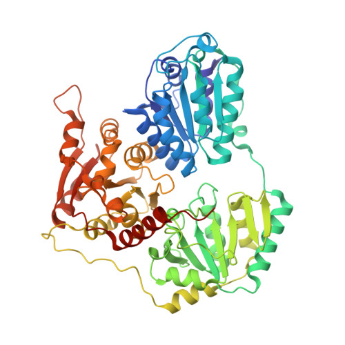
Structural Insights Into the Pre-Reaction State of Pyruvate Decarboxylase from Zymomonas Mobilis

Pei, X.Y.Erixon, K.Luisi, B.F.Leeper, F.J.

(2010) Biochemistry 49: 1727

  • DOI: https://doi.org/10.1021/bi901864j
  • Primary Citation of Related Structures:  
    2WVA, 2WVG, 2WVH

  • PubMed Abstract: 

    Pyruvate decarboxylase (PDC) uses thiamine diphosphate as an essential cofactor to catalyze the formation of acetaldehyde on the pathway of ethanol synthesis. Here we report the crystallographic image of a prereaction intermediate of a bacterial pyruvate decarboxylase prepared by cocrystallizing the enzyme with pyruvate and a stable analogue of the cofactor's activated ylid form. A second crystal structure of PDC in complex with fluoride shows that the ion organizes a water molecule that occludes the pyruvate binding site, accounting for the inhibitory effect of the halide. Also reported is a structure of the cofactor-free apo form, which when compared to the structure of the holo form indicates how thiamine diphosphate organizes the active site pocket of pyruvate decarboxylase to support catalysis. Guided by the structural and enzymatic data, we propose roles for several key residues in the catalytic mechanism.


  • Organizational Affiliation

    Department of Biochemistry, University of Cambridge, Tennis Court Road, Cambridge CB2 1QA, UK.


Macromolecules
Find similar proteins by:  (by identity cutoff)  |  3D Structure
Entity ID: 1
MoleculeChains Sequence LengthOrganismDetailsImage
PYRUVATE DECARBOXYLASEA,
B,
C [auth E],
D [auth F]
568Zymomonas mobilisMutation(s): 0 
EC: 4.1.1.1
UniProt
Find proteins for P06672 (Zymomonas mobilis subsp. mobilis (strain ATCC 31821 / ZM4 / CP4))
Explore P06672 
Go to UniProtKB:  P06672
Entity Groups  
Sequence Clusters30% Identity50% Identity70% Identity90% Identity95% Identity100% Identity
UniProt GroupP06672
Sequence Annotations
Expand
  • Reference Sequence
Small Molecules
Ligands 3 Unique
IDChains Name / Formula / InChI Key2D Diagram3D Interactions
TPU
Query on TPU

Download Ideal Coordinates CCD File 
F [auth A],
I [auth B],
L [auth E],
P [auth F]
2-{1-[(4-AMINO-2-METHYLPYRIMIDIN-5-YL)METHYL]-5-METHYL-1H-1,2,3-TRIAZOL-4-YL}ETHYL TRIHYDROGEN DIPHOSPHATE
C11 H18 N6 O7 P2
WHMXQEYVWLIXHH-UHFFFAOYSA-N
MG
Query on MG

Download Ideal Coordinates CCD File 
G [auth A],
J [auth B],
M [auth E],
N [auth F]
MAGNESIUM ION
Mg
JLVVSXFLKOJNIY-UHFFFAOYSA-N
F
Query on F

Download Ideal Coordinates CCD File 
E [auth A],
H [auth B],
K [auth E],
O [auth F]
FLUORIDE ION
F
KRHYYFGTRYWZRS-UHFFFAOYSA-M
Experimental Data & Validation

Experimental Data

  • Method: X-RAY DIFFRACTION
  • Resolution: 1.75 Å
  • R-Value Free: 0.187 
  • R-Value Work: 0.157 
  • R-Value Observed: 0.158 
  • Space Group: P 1
Unit Cell:
Length ( Å )Angle ( ˚ )
a = 70.277α = 73.44
b = 92.092β = 85.76
c = 98.63γ = 67.82
Software Package:
Software NamePurpose
PHENIXrefinement
MOSFLMdata reduction
SCALAdata scaling
TRUNCATEdata scaling
AMoREphasing

Structure Validation

View Full Validation Report


Ligand Structure Quality Assessment 


Entry History 

Deposition Data

Revision History  (Full details and data files)

  • Version 1.0: 2010-02-09
    Type: Initial release
  • Version 1.1: 2011-10-19
    Changes: Database references, Derived calculations, Refinement description, Version format compliance
  • Version 1.2: 2019-03-06
    Changes: Data collection, Experimental preparation, Other
  • Version 1.3: 2023-12-20
    Changes: Data collection, Database references, Derived calculations, Other, Refinement description