2WUH | pdb_00002wuh

Crystal structure of the DDR2 discoidin domain bound to a triple- helical collagen peptide


Experimental Data Snapshot

  • Method: X-RAY DIFFRACTION
  • Resolution: 1.60 Å
  • R-Value Free: 
    0.230 (Depositor), 0.224 (DCC) 
  • R-Value Work: 
    0.202 (Depositor), 0.197 (DCC) 
  • R-Value Observed: 
    0.202 (Depositor) 

Starting Models: experimental
View more details

wwPDB Validation 3D Report Full Report

Validation slider image for 2WUH

This is version 1.3 of the entry. See complete history

Literature

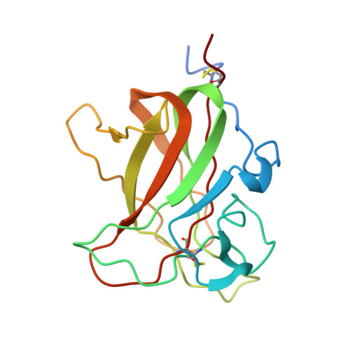
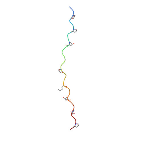
Crystallographic Insight Into Collagen Recognition by Discoidin Domain Receptor 2

Carafoli, F.Bihan, D.Stathopoulos, S.Konitsiotis, A.D.Kvansakul, M.Farndale, R.W.Leitinger, B.Hohenester, E.

(2009) Structure 17: 1573

  • DOI: https://doi.org/10.1016/j.str.2009.10.012
  • Primary Citation Related Structures: 
    2WUH

  • PubMed Abstract: 

    The discoidin domain receptors, DDR1 and DDR2, are widely expressed receptor tyrosine kinases that are activated by triple-helical collagen. They control important aspects of cell behavior and are dysregulated in several human diseases. The major DDR2-binding site in collagens I-III is a GVMGFO motif (O is hydroxyproline) that also binds the matricellular protein SPARC. We have determined the crystal structure of the discoidin domain of human DDR2 bound to a triple-helical collagen peptide. The GVMGFO motifs of two collagen chains are recognized by an amphiphilic pocket delimited by a functionally critical tryptophan residue and a buried salt bridge. Collagen binding results in structural changes of DDR2 surface loops that may be linked to the process of receptor activation. A comparison of the GVMGFO-binding sites of DDR2 and SPARC reveals a striking case of convergent evolution in collagen recognition.


  • Organizational Affiliation
    • Department of Life Sciences, Imperial College London, London SW7 2AZ, UK.

Macromolecule Content 

  • Total Structure Weight: 28.01 kDa 
  • Atom Count: 2,021 
  • Modeled Residue Count: 249 
  • Deposited Residue Count: 265 
  • Unique protein chains: 2

Macromolecules

Find similar proteins by:|  3D Structure
Entity ID: 1
MoleculeChains  Sequence LengthOrganismDetailsImage
DISCOIDIN DOMAIN RECEPTOR 2178Homo sapiensMutation(s): 0 
EC: 2.7.10.1
Membrane Entity: Yes 
UniProt & NIH Common Fund Data Resources
Find proteins for Q16832 (Homo sapiens)
Explore Q16832 
Go to UniProtKB:  Q16832
PHAROS:  Q16832
GTEx:  ENSG00000162733 
Entity Groups
Sequence Clusters30% Identity50% Identity70% Identity90% Identity95% Identity100% Identity
UniProt GroupQ16832
Sequence Annotations
Expand
Reference Sequence
Find similar proteins by:|  3D Structure
Entity ID: 2
MoleculeChains  Sequence LengthOrganismDetailsImage
COLLAGEN PEPTIDE
B, C, D
29Homo sapiensMutation(s): 0 

Small Molecules

Modified Residues  2 Unique
IDChains TypeFormula2D DiagramParent
HYP
Query on HYP
B, C, D
L-PEPTIDE LINKINGC5 H9 N O3PRO
NLE
Query on NLE
B, C, D
L-PEPTIDE LINKINGC6 H13 N O2LEU

Experimental Data & Validation

Experimental Data

  • Method: X-RAY DIFFRACTION
  • Resolution: 1.60 Å
  • R-Value Free:  0.230 (Depositor), 0.224 (DCC) 
  • R-Value Work:  0.202 (Depositor), 0.197 (DCC) 
  • R-Value Observed: 0.202 (Depositor) 
Space Group: P 1
Unit Cell:
Length ( Å )Angle ( ˚ )
a = 33.397α = 66.19
b = 40.863β = 88.9
c = 48.917γ = 76.92
Software Package:
Software NamePurpose
CNSrefinement
MOSFLMdata reduction
SCALAdata scaling
PHASERphasing

Structure Validation

View Full Validation Report


Entry History 

Deposition Data

Revision History  (Full details and data files)

  • Version 1.0: 2009-12-29
    Type: Initial release
  • Version 1.1: 2011-05-08
    Changes: Version format compliance
  • Version 1.2: 2011-07-13
    Changes: Version format compliance
  • Version 1.3: 2023-12-20
    Changes: Data collection, Database references, Derived calculations, Other, Refinement description