2WTZ | pdb_00002wtz

MurE ligase of Mycobacterium Tuberculosis


Experimental Data Snapshot

  • Method: X-RAY DIFFRACTION
  • Resolution: 3.00 Å
  • R-Value Free: 
    0.251 (Depositor), 0.248 (DCC) 
  • R-Value Work: 
    0.193 (Depositor), 0.196 (DCC) 
  • R-Value Observed: 
    0.195 (Depositor) 

Starting Model: experimental
View more details

wwPDB Validation 3D Report Full Report

Validation slider image for 2WTZ

Ligand Structure Quality Assessment 


This is version 1.2 of the entry. See complete history

Literature

ATP-Dependent Mure Ligase in Mycobacterium Tuberculosis: Biochemical and Structural Characterisation.

Basavannacharya, C.Robertson, G.Munshi, T.Keep, N.H.Bhakta, S.

(2010) Tuberculosis (Edinb) 90: 16

  • DOI: https://doi.org/10.1016/j.tube.2009.10.007
  • Primary Citation Related Structures: 
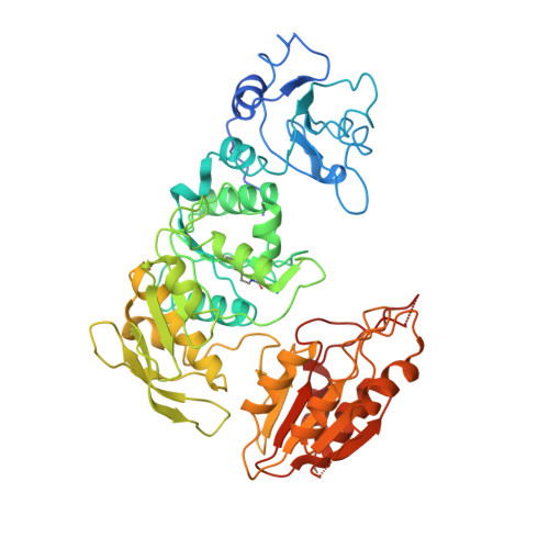
    2WTZ

  • PubMed Abstract: 

    New therapies are required against Mycobacterium tuberculosis and its cell wall peptidoglycan biosynthesis is a potential therapeutic target. UDP-MurNAc-tripeptide ligase (MurE) is a member of the ATP-dependent ligase family, which incorporate amino acids including meso-diaminopimelic acid (m-DAP) into peptidoglycan during synthesis in a species-specific manner. In the present study, we have cloned, over-expressed, and characterised MurE from M. tuberculosis (Mtb-MurE). The crystal structure has been determined at 3.0A resolution in the presence of the substrate UDP-MurNAc-l-Ala-d-Glu (UAG). The activity of the enzyme was measured through estimating inorganic phosphate released in a non-radioactive high-throughput colourimetric assay. UDP-MurNAc-l-Ala-d-Glu-m-DAP (UMT) formation coupled to inorganic phosphate release was confirmed by HPLC and mass spectrometric analyses. Kinetic constants were determined for a range of natural substrates using optimised conditions. From our findings, it is evident that Mtb-MurE is highly specific in adding m-DAP to UDP-MurNAc-dipeptide and ATP-hydrolysis is an absolute requirement for its activity.


  • Organizational Affiliation
    • Institute of Structural and Molecular Biology, Department of Biological Sciences, Birkbeck, University of London, Malet Street, London WC1E 7HX, UK.

Macromolecule Content 

  • Total Structure Weight: 225.43 kDa 
  • Atom Count: 14,008 
  • Modeled Residue Count: 1,919 
  • Deposited Residue Count: 2,140 
  • Unique protein chains: 1

Macromolecules

Find similar proteins by:|  3D Structure
Entity ID: 1
MoleculeChains  Sequence LengthOrganismDetailsImage
UDP-N-ACETYLMURAMOYL-L-ALANYL-D-GLUTAMATE--2,6-DIAMINOPIMELATE LIGASE
A, B, C, D
535Mycobacterium tuberculosis H37RvMutation(s): 0 
EC: 6.3.2.13
UniProt
Find proteins for P9WJL3 (Mycobacterium tuberculosis (strain ATCC 25618 / H37Rv))
Explore P9WJL3 
Go to UniProtKB:  P9WJL3
Entity Groups
Sequence Clusters30% Identity50% Identity70% Identity90% Identity95% Identity100% Identity
UniProt GroupP9WJL3
Sequence Annotations
Expand
Reference Sequence

Small Molecules

Ligands 2 Unique
IDChains Name / Formula / InChI Key2D Diagram3D Interactions
UAG

Query on UAG


Download:Ideal Coordinates CCD File
E [auth A],
G [auth B],
I [auth C],
K [auth D]
URIDINE-5'-DIPHOSPHATE-N-ACETYLMURAMOYL-L-ALANINE-D-GLUTAMATE
C28 H43 N5 O23 P2
OJZCATPXPWFLHF-HPUCEMLMSA-N
MG

Query on MG


Download:Ideal Coordinates CCD File
F [auth A],
H [auth B],
J [auth C],
L [auth D]
MAGNESIUM ION
Mg
JLVVSXFLKOJNIY-UHFFFAOYSA-N
Modified Residues  1 Unique
IDChains TypeFormula2D DiagramParent
KCX
Query on KCX
A, B, C, D
L-PEPTIDE LINKINGC7 H14 N2 O4LYS

Experimental Data & Validation

Experimental Data

  • Method: X-RAY DIFFRACTION
  • Resolution: 3.00 Å
  • R-Value Free:  0.251 (Depositor), 0.248 (DCC) 
  • R-Value Work:  0.193 (Depositor), 0.196 (DCC) 
  • R-Value Observed: 0.195 (Depositor) 
Space Group: P 1
Unit Cell:
Length ( Å )Angle ( ˚ )
a = 76.92α = 111.09
b = 79.8β = 92.16
c = 82.92γ = 93.98
Software Package:
Software NamePurpose
REFMACrefinement
MOSFLMdata reduction
SCALAdata scaling
MrBUMPphasing

Structure Validation

View Full Validation Report


Ligand Structure Quality Assessment 


Entry History 

Deposition Data

Revision History  (Full details and data files)

  • Version 1.0: 2009-12-15
    Type: Initial release
  • Version 1.1: 2011-07-13
    Changes: Advisory, Version format compliance
  • Version 1.2: 2023-12-20
    Changes: Data collection, Database references, Derived calculations, Other, Refinement description