2WTV

Aurora-A Inhibitor Structure


Experimental Data Snapshot

  • Method: X-RAY DIFFRACTION
  • Resolution: 2.40 Å
  • R-Value Free: 0.211 
  • R-Value Work: 0.164 
  • R-Value Observed: 0.167 

wwPDB Validation   3D Report Full Report


Ligand Structure Quality Assessment 


This is version 1.3 of the entry. See complete history


Literature

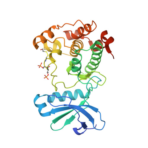
Crystal Structure of an Aurora-A Mutant that Mimics Aurora-B Bound to Mln8054: Insights Into Selectivity and Drug Design.

Dodson, C.A.Kosmopoulou, M.Richards, M.W.Atrash, B.Bavetsias, V.Blagg, J.Bayliss, R.

(2010) Biochem J 427: 19

  • DOI: https://doi.org/10.1042/BJ20091530
  • Primary Citation of Related Structures:  
    2WTV, 2WTW

  • PubMed Abstract: 

    The production of selective protein kinase inhibitors is often frustrated by the similarity of the enzyme active sites. For this reason, it is challenging to design inhibitors that discriminate between the three Aurora kinases, which are important targets in cancer drug discovery. We have used a triple-point mutant of Aurora-A (AurAx3) which mimics the active site of Aurora-B to investigate the structural basis of MLN8054 selectivity. The bias toward Aurora-A inhibition by MLN8054 is fully recapitulated by AurAx3 in vitro. X-ray crystal structures of the complex suggest that the basis for the discrimination is electrostatic repulsion due to the T217E substitution, which we have confirmed using a single-point mutant. The activation loop of Aurora-A in the AurAx3-MLN8054 complex exhibits an unusual conformation in which Asp274 and Phe275 side chains point into the interior of the protein. There is to our knowledge no documented precedent for this conformation, which we have termed DFG-up. The sequence requirements of the DFG-up conformation suggest that it might be accessible to only a fraction of kinases. MLN8054 thus circumvents the problem of highly homologous active sites. Binding of MLN8054 to Aurora-A switches the character of a pocket within the active site from polar to a hydrophobic pocket, similar to what is observed in the structure of Aurora-A bound to a compound that induces DFG-out. We propose that targeting this pocket may be a productive route in the design of selective kinase inhibitors and describe the structural basis for the rational design of these compounds.


  • Organizational Affiliation

    Section of Structural Biology, The Institute of Cancer Research, Chester Beatty Laboratories, 237 Fulham Road, London SW36JB, U.K.


Macromolecules
Find similar proteins by:  (by identity cutoff)  |  3D Structure
Entity ID: 1
MoleculeChains Sequence LengthOrganismDetailsImage
SERINE/THREONINE-PROTEIN KINASE 6 AURORA/IPL1-RELATED KINASE 1, BREAST TUMOR-AMPLIFIED KINASE, AURORA-A, AURORA-RELATED KINASE 1, HARK1
A, B, C, D
285Homo sapiensMutation(s): 3 
EC: 2.7.11.1
UniProt & NIH Common Fund Data Resources
Find proteins for O14965 (Homo sapiens)
Explore O14965 
Go to UniProtKB:  O14965
PHAROS:  O14965
GTEx:  ENSG00000087586 
Entity Groups  
Sequence Clusters30% Identity50% Identity70% Identity90% Identity95% Identity100% Identity
UniProt GroupO14965
Sequence Annotations
Expand
  • Reference Sequence
Small Molecules
Ligands 4 Unique
IDChains Name / Formula / InChI Key2D Diagram3D Interactions
ZZL
Query on ZZL

Download Ideal Coordinates CCD File 
AA [auth D],
E [auth A],
J [auth B],
W [auth C]
4-{[9-CHLORO-7-(2,6-DIFLUOROPHENYL)-5H-PYRIMIDO[5,4-D][2]BENZAZEPIN-2-YL]AMINO}BENZOIC ACID
C25 H15 Cl F2 N4 O2
HHFBDROWDBDFBR-UHFFFAOYSA-N
DMS
Query on DMS

Download Ideal Coordinates CCD File 
I [auth A],
K [auth B]
DIMETHYL SULFOXIDE
C2 H6 O S
IAZDPXIOMUYVGZ-UHFFFAOYSA-N
EDO
Query on EDO

Download Ideal Coordinates CCD File 
BA [auth D]
F [auth A]
N [auth B]
O [auth B]
P [auth B]
BA [auth D],
F [auth A],
N [auth B],
O [auth B],
P [auth B],
Q [auth B],
R [auth B],
S [auth B],
U [auth C],
V [auth C],
Z [auth C]
1,2-ETHANEDIOL
C2 H6 O2
LYCAIKOWRPUZTN-UHFFFAOYSA-N
ACT
Query on ACT

Download Ideal Coordinates CCD File 
CA [auth D]
DA [auth D]
G [auth A]
H [auth A]
L [auth B]
CA [auth D],
DA [auth D],
G [auth A],
H [auth A],
L [auth B],
M [auth B],
T [auth B],
X [auth C],
Y [auth C]
ACETATE ION
C2 H3 O2
QTBSBXVTEAMEQO-UHFFFAOYSA-M
Modified Residues  2 Unique
IDChains TypeFormula2D DiagramParent
CME
Query on CME
A, B, C, D
L-PEPTIDE LINKINGC5 H11 N O3 S2CYS
TPO
Query on TPO
A, B, C, D
L-PEPTIDE LINKINGC4 H10 N O6 PTHR
Binding Affinity Annotations 
IDSourceBinding Affinity
ZZL PDBBind:  2WTV Ki: 1.8 (nM) from 1 assay(s)
BindingDB:  2WTV Ki: 7 (nM) from 1 assay(s)
Kd: 6.5 (nM) from 1 assay(s)
IC50: min: 1, max: 34 (nM) from 8 assay(s)
Binding MOAD:  2WTV Ki: 1.8 (nM) from 1 assay(s)
Experimental Data & Validation

Experimental Data

  • Method: X-RAY DIFFRACTION
  • Resolution: 2.40 Å
  • R-Value Free: 0.211 
  • R-Value Work: 0.164 
  • R-Value Observed: 0.167 
  • Space Group: P 31
Unit Cell:
Length ( Å )Angle ( ˚ )
a = 118.707α = 90
b = 118.707β = 90
c = 135.89γ = 120
Software Package:
Software NamePurpose
PHENIXrefinement
MOSFLMdata reduction
SCALAdata scaling
PHASERphasing

Structure Validation

View Full Validation Report


Ligand Structure Quality Assessment 


Entry History 

Deposition Data

Revision History  (Full details and data files)

  • Version 1.0: 2010-01-26
    Type: Initial release
  • Version 1.1: 2011-05-08
    Changes: Version format compliance
  • Version 1.2: 2011-07-13
    Changes: Version format compliance
  • Version 1.3: 2023-12-20
    Changes: Data collection, Database references, Derived calculations, Other, Refinement description