2WTD | pdb_00002wtd

Crystal structure of Chk2 in complex with an inhibitor


Experimental Data Snapshot

  • Method: X-RAY DIFFRACTION
  • Resolution: 2.75 Å
  • R-Value Free: 
    0.258 (Depositor), 0.258 (DCC) 
  • R-Value Work: 
    0.191 (Depositor), 0.189 (DCC) 
  • R-Value Observed: 
    0.194 (Depositor) 

Starting Model: experimental
View more details

wwPDB Validation 3D Report Full Report

Validation slider image for 2WTD

Ligand Structure Quality Assessment 


This is version 1.3 of the entry. See complete history

Literature

Identification and Characterisation of 2-Aminopyridine Inhibitors of Checkpoint Kinase 2

Hilton, S.Naud, S.Caldwell, J.J.Boxall, K.Burns, S.Anderson, V.E.Antoni, L.Allen, C.E.Pearl, L.H.Oliver, A.W.Aherne, G.W.Garrett, M.D.Collins, I.

(2010) Bioorg Med Chem 18: 707

  • DOI: https://doi.org/10.1016/j.bmc.2009.11.058
  • Primary Citation Related Structures: 
    2WTC, 2WTD, 2WTI, 2WTJ

  • PubMed Abstract: 

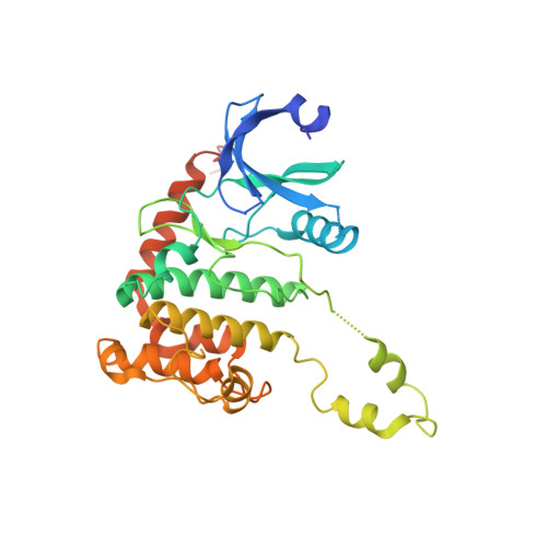
    5-(Hetero)aryl-3-(4-carboxamidophenyl)-2-aminopyridine inhibitors of CHK2 were identified from high throughput screening of a kinase-focussed compound library. Rapid exploration of the hits through straightforward chemistry established structure-activity relationships and a proposed ATP-competitive binding mode which was verified by X-ray crystallography of several analogues bound to CHK2. Variation of the 5-(hetero)aryl substituent identified bicyclic dioxolane and dioxane groups which improved the affinity and the selectivity of the compounds for CHK2 versus CHK1. The 3-(4-carboxamidophenyl) substituent could be successfully replaced by acyclic omega-aminoalkylamides, which made additional polar interactions within the binding site and led to more potent inhibitors of CHK2. Compounds from this series showed activity in cell-based mechanistic assays for inhibition of CHK2.


  • Organizational Affiliation
    • Cancer Research UK Centre for Cancer Therapeutics, The Institute of Cancer Research, 15 Cotswold Road, Sutton, Surrey SM2 5NG, UK.

Macromolecule Content 

  • Total Structure Weight: 37.69 kDa 
  • Atom Count: 2,357 
  • Modeled Residue Count: 285 
  • Deposited Residue Count: 329 
  • Unique protein chains: 1

Macromolecules

Find similar proteins by:|  3D Structure
Entity ID: 1
MoleculeChains  Sequence LengthOrganismDetailsImage
SERINE/THREONINE-PROTEIN KINASE CHK2329Homo sapiensMutation(s): 0 
EC: 2.7.11.1
UniProt & NIH Common Fund Data Resources
Find proteins for O96017 (Homo sapiens)
Explore O96017 
Go to UniProtKB:  O96017
PHAROS:  O96017
GTEx:  ENSG00000183765 
Entity Groups
Sequence Clusters30% Identity50% Identity70% Identity90% Identity95% Identity100% Identity
UniProt GroupO96017
Sequence Annotations
Expand
Reference Sequence

Experimental Data & Validation

Experimental Data

  • Method: X-RAY DIFFRACTION
  • Resolution: 2.75 Å
  • R-Value Free:  0.258 (Depositor), 0.258 (DCC) 
  • R-Value Work:  0.191 (Depositor), 0.189 (DCC) 
  • R-Value Observed: 0.194 (Depositor) 
Space Group: P 32 2 1
Unit Cell:
Length ( Å )Angle ( ˚ )
a = 90.89α = 90
b = 90.89β = 90
c = 92.83γ = 120
Software Package:
Software NamePurpose
PHENIXrefinement
MOSFLMdata reduction
SCALAdata scaling
PHASERphasing

Structure Validation

View Full Validation Report


Ligand Structure Quality Assessment 


Entry History 

Revision History  (Full details and data files)

  • Version 1.0: 2009-12-29
    Type: Initial release
  • Version 1.1: 2011-05-08
    Changes: Version format compliance
  • Version 1.2: 2011-07-13
    Changes: Version format compliance
  • Version 1.3: 2023-12-20
    Changes: Data collection, Database references, Derived calculations, Other, Refinement description