2WRW | pdb_00002wrw

Semi-synthetic highly active analogue of human insulin D-ProB26-DTI- NH2


Experimental Data Snapshot

  • Method: X-RAY DIFFRACTION
  • Resolution: 2.41 Å
  • R-Value Free: 
    0.301 (Depositor), 0.282 (DCC) 
  • R-Value Work: 
    0.237 (Depositor), 0.247 (DCC) 
  • R-Value Observed: 
    0.239 (Depositor) 

Starting Model: experimental
View more details

wwPDB Validation 3D Report Full Report

Validation slider image for 2WRW

This is version 1.3 of the entry. See complete history

Literature

Implications for the Active Form of Human Insulin Based on the Structural Convergence of Highly Active Hormone Analogues.

Jiracek, J.Zakova, L.Antolikova, E.Watson, C.J.Turkenburg, J.P.Dodson, G.G.Brzozowski, A.M.

(2010) Proc Natl Acad Sci U S A 107: 1966

  • DOI: https://doi.org/10.1073/pnas.0911785107
  • Primary Citation Related Structures: 
    2WRU, 2WRV, 2WRW, 2WRX, 2WS0, 2WS1, 2WS4, 2WS6, 2WS7

  • PubMed Abstract: 

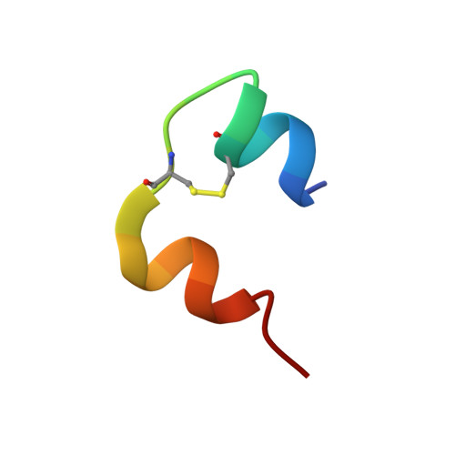
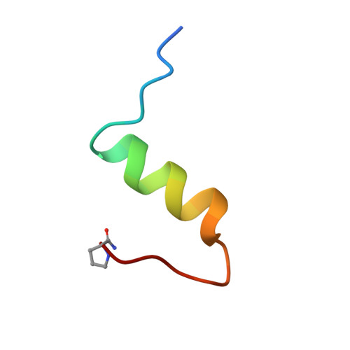
    Insulin is a key protein hormone that regulates blood glucose levels and, thus, has widespread impact on lipid and protein metabolism. Insulin action is manifested through binding of its monomeric form to the Insulin Receptor (IR). At present, however, our knowledge about the structural behavior of insulin is based upon inactive, multimeric, and storage-like states. The active monomeric structure, when in complex with the receptor, must be different as the residues crucial for the interactions are buried within the multimeric forms. Although the exact nature of the insulin's induced-fit is unknown, there is strong evidence that the C-terminal part of the B-chain is a dynamic element in insulin activation and receptor binding. Here, we present the design and analysis of highly active (200-500%) insulin analogues that are truncated at residue 26 of the B-chain (B(26)). They show a structural convergence in the form of a new beta-turn at B(24)-B(26). We propose that the key element in insulin's transition, from an inactive to an active state, may be the formation of the beta-turn at B(24)-B(26) associated with a trans to cis isomerisation at the B(25)-B(26) peptide bond. Here, this turn is achieved with N-methylated L-amino acids adjacent to the trans to cis switch at the B(25)-B(26) peptide bond or by the insertion of certain D-amino acids at B(26). The resultant conformational changes unmask previously buried amino acids that are implicated in IR binding and provide structural details for new approaches in rational design of ligands effective in combating diabetes.


  • Organizational Affiliation
    • Institute of Organic Chemistry and Biochemistry, Academy of Sciences of the Czech Republic, vvi, Flemingovo nám 2, 166 10 Prague 6, Czech Republic.

Macromolecule Content 

  • Total Structure Weight: 5.32 kDa 
  • Atom Count: 384 
  • Modeled Residue Count: 46 
  • Deposited Residue Count: 47 
  • Unique protein chains: 2

Macromolecules

Find similar proteins by:  Sequence   |   3D Structure  
Entity ID: 1
MoleculeChains  Sequence LengthOrganismDetailsImage
INSULIN A CHAIN21Homo sapiensMutation(s): 0 
UniProt & NIH Common Fund Data Resources
Find proteins for P01308 (Homo sapiens)
Explore P01308 
Go to UniProtKB:  P01308
PHAROS:  P01308
GTEx:  ENSG00000254647 
Entity Groups
Sequence Clusters30% Identity50% Identity70% Identity90% Identity95% Identity100% Identity
UniProt GroupP01308
Sequence Annotations
Expand
Reference Sequence
Find similar proteins by:|  3D Structure
Entity ID: 2
MoleculeChains  Sequence LengthOrganismDetailsImage
INSULIN B CHAIN26Homo sapiensMutation(s): 1 
UniProt & NIH Common Fund Data Resources
Find proteins for P01308 (Homo sapiens)
Explore P01308 
Go to UniProtKB:  P01308
PHAROS:  P01308
GTEx:  ENSG00000254647 
Entity Groups
Sequence Clusters30% Identity50% Identity70% Identity90% Identity95% Identity100% Identity
UniProt GroupP01308
Sequence Annotations
Expand
Reference Sequence

Small Molecules

Modified Residues  1 Unique
IDChains TypeFormula2D DiagramParent
PR9
Query on PR9
B
D-PEPTIDE LINKINGC5 H10 N2 OPRO

Experimental Data & Validation

Experimental Data

  • Method: X-RAY DIFFRACTION
  • Resolution: 2.41 Å
  • R-Value Free:  0.301 (Depositor), 0.282 (DCC) 
  • R-Value Work:  0.237 (Depositor), 0.247 (DCC) 
  • R-Value Observed: 0.239 (Depositor) 
Space Group: I 41 2 2
Unit Cell:
Length ( Å )Angle ( ˚ )
a = 39.372α = 90
b = 39.372β = 90
c = 123.928γ = 90
Software Package:
Software NamePurpose
REFMACrefinement
DENZOdata reduction
SCALEPACKdata scaling
MOLREPphasing

Structure Validation

View Full Validation Report


Entry History 

Deposition Data

Revision History  (Full details and data files)

  • Version 1.0: 2010-02-09
    Type: Initial release
  • Version 1.1: 2011-07-13
    Changes: Advisory, Version format compliance
  • Version 1.2: 2012-06-06
    Changes: Other
  • Version 1.3: 2023-12-20
    Changes: Data collection, Database references, Derived calculations, Other, Refinement description