2WJY | pdb_00002wjy

Crystal structure of the complex between human nonsense mediated decay factors UPF1 and UPF2 Orthorhombic form


Experimental Data Snapshot

  • Method: X-RAY DIFFRACTION
  • Resolution: 2.50 Å
  • R-Value Free: 
    0.240 (Depositor), 0.246 (DCC) 
  • R-Value Work: 
    0.196 (Depositor), 0.202 (DCC) 
  • R-Value Observed: 
    0.198 (Depositor) 

Starting Models: experimental
View more details

wwPDB Validation 3D Report Full Report

Validation slider image for 2WJY

This is version 1.2 of the entry. See complete history

Literature

Unusual Bipartite Mode of Interaction between the Nonsense-Mediated Decay Factors, Upf1 and Upf2.

Clerici, M.Mourao, A.Gutsche, I.Gehring, N.H.Hentze, M.W.Kulozik, A.Kadlec, J.Sattler, M.Cusack, S.

(2009) EMBO J 28: 2293

  • DOI: https://doi.org/10.1038/emboj.2009.175
  • Primary Citation Related Structures: 
    2WJV, 2WJY

  • PubMed Abstract: 

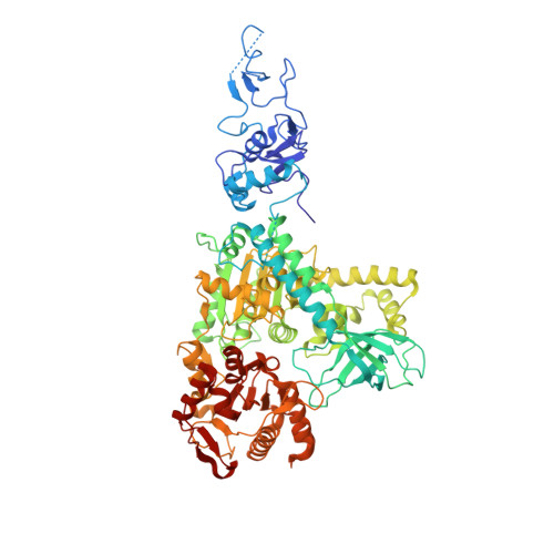
    Nonsense-mediated decay (NMD) is a eukaryotic quality control mechanism that degrades mRNAs carrying premature stop codons. In mammalian cells, NMD is triggered when UPF2 bound to UPF3 on a downstream exon junction complex interacts with UPF1 bound to a stalled ribosome. We report structural studies on the interaction between the C-terminal region of UPF2 and intact UPF1. Crystal structures, confirmed by EM and SAXS, show that the UPF1 CH-domain is docked onto its helicase domain in a fixed configuration. The C-terminal region of UPF2 is natively unfolded but binds through separated alpha-helical and beta-hairpin elements to the UPF1 CH-domain. The alpha-helical region binds sixfold more weakly than the beta-hairpin, whereas the combined elements bind 80-fold more tightly. Cellular assays show that NMD is severely affected by mutations disrupting the beta-hairpin binding, but not by those only affecting alpha-helix binding. We propose that the bipartite mode of UPF2 binding to UPF1 brings the ribosome and the EJC in close proximity by forming a tight complex after an initial weak encounter with either element.


  • Organizational Affiliation
    • European Molecular Biology Laboratory, Grenoble Outstation, Grenoble Cedex 9, France.

Macromolecule Content 

  • Total Structure Weight: 90.95 kDa 
  • Atom Count: 6,465 
  • Modeled Residue Count: 773 
  • Deposited Residue Count: 800 
  • Unique protein chains: 1

Macromolecules

Find similar proteins by:|  3D Structure
Entity ID: 1
MoleculeChains  Sequence LengthOrganismDetailsImage
REGULATOR OF NONSENSE TRANSCRIPTS 1800Homo sapiensMutation(s): 0 
EC: 3.6.1 (PDB Primary Data), 3.6.4.13 (UniProt), 3.6.4.12 (UniProt)
UniProt & NIH Common Fund Data Resources
Find proteins for Q92900 (Homo sapiens)
Explore Q92900 
Go to UniProtKB:  Q92900
PHAROS:  Q92900
GTEx:  ENSG00000005007 
Entity Groups
Sequence Clusters30% Identity50% Identity70% Identity90% Identity95% Identity100% Identity
UniProt GroupQ92900
Sequence Annotations
Expand
Reference Sequence

Small Molecules

Ligands 2 Unique
IDChains Name / Formula / InChI Key2D Diagram3D Interactions
SO4

Query on SO4


Download:Ideal Coordinates CCD File
E [auth A]
F [auth A]
G [auth A]
H [auth A]
I [auth A]
E [auth A],
F [auth A],
G [auth A],
H [auth A],
I [auth A],
J [auth A],
K [auth A],
L [auth A]
SULFATE ION
O4 S
QAOWNCQODCNURD-UHFFFAOYSA-L
ZN

Query on ZN


Download:Ideal Coordinates CCD File
B [auth A],
C [auth A],
D [auth A]
ZINC ION
Zn
PTFCDOFLOPIGGS-UHFFFAOYSA-N

Experimental Data & Validation

Experimental Data

  • Method: X-RAY DIFFRACTION
  • Resolution: 2.50 Å
  • R-Value Free:  0.240 (Depositor), 0.246 (DCC) 
  • R-Value Work:  0.196 (Depositor), 0.202 (DCC) 
  • R-Value Observed: 0.198 (Depositor) 
Space Group: I 2 2 2
Unit Cell:
Length ( Å )Angle ( ˚ )
a = 64.91α = 90
b = 129.14β = 90
c = 311.27γ = 90
Software Package:
Software NamePurpose
REFMACrefinement
XDSdata reduction
XSCALEdata scaling
PHASERphasing

Structure Validation

View Full Validation Report


Entry History 

Deposition Data

Revision History  (Full details and data files)

  • Version 1.0: 2009-07-07
    Type: Initial release
  • Version 1.1: 2011-07-13
    Changes: Advisory, Version format compliance
  • Version 1.2: 2023-12-13
    Changes: Data collection, Database references, Derived calculations, Other, Refinement description