2WJS | pdb_00002wjs

Crystal structure of the LG1-3 region of the laminin alpha2 chain


Experimental Data Snapshot

  • Method: X-RAY DIFFRACTION
  • Resolution: 2.80 Å
  • R-Value Free: 
    0.266 (Depositor), 0.258 (DCC) 
  • R-Value Work: 
    0.212 (Depositor), 0.206 (DCC) 
  • R-Value Observed: 
    0.212 (Depositor) 

Starting Model: experimental
View more details

wwPDB Validation 3D Report Full Report

Validation slider image for 2WJS

Ligand Structure Quality Assessment 


This is version 1.5 of the entry. See complete history

Literature

Crystal Structure of the Lg1-3 Region of the Laminin {Alpha}2 Chain.

Carafoli, F.Clout, N.J.Hohenester, E.

(2009) J Biological Chem 284: 22786

  • DOI: https://doi.org/10.1074/jbc.M109.026658
  • Primary Citation Related Structures: 
    2WJS

  • PubMed Abstract: 

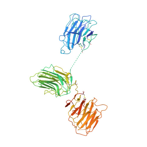
    Laminins are large heterotrimeric glycoproteins with many essential functions in basement membrane assembly and function. Cell adhesion to laminins is mediated by a tandem of five laminin G-like (LG) domains at the C terminus of the alpha chain. Integrin binding requires an intact LG1-3 region, as well as contributions from the coiled coil formed by the alpha, beta, and gamma chains. We have determined the crystal structure at 2.8-A resolution of the LG1-3 region of the laminin alpha2 chain (alpha 2LG1-3). The three LG domains adopt typical beta-sandwich folds, with canonical calcium binding sites in LG1 and LG2. LG2 and LG3 interact through a substantial interface, but LG1 is completely dissociated from the LG2-3 pair. We suggest that the missing gamma chain tail may be required to stabilize the interaction between LG1 and LG2-3 in the biologically active conformation. A global analysis of N-linked glycosylation sites shows that the beta-sandwich faces of LG1 are free of carbohydrate modifications in all five laminin alpha chains, suggesting that these surfaces may harbor the integrin binding site. The alpha 2LG1-3 structure provides the first atomic view of the integrin binding region of laminins.


  • Organizational Affiliation
    • Department of Life Sciences, Imperial College London, London SW7 2AZ, United Kingdom.

Macromolecule Content 

  • Total Structure Weight: 68.18 kDa 
  • Atom Count: 4,056 
  • Modeled Residue Count: 520 
  • Deposited Residue Count: 608 
  • Unique protein chains: 1

Macromolecules

Find similar proteins by:|  3D Structure
Entity ID: 1
MoleculeChains  Sequence LengthOrganismDetailsImage
LAMININ SUBUNIT ALPHA-2608Mus musculusMutation(s): 0 
UniProt
Find proteins for Q60675 (Mus musculus)
Explore Q60675 
Go to UniProtKB:  Q60675
Entity Groups
Sequence Clusters30% Identity50% Identity70% Identity90% Identity95% Identity100% Identity
UniProt GroupQ60675
Glycosylation
Glycosylation Sites: 4Go to GlyGen: Q60675-1
Sequence Annotations
Expand
Reference Sequence

Experimental Data & Validation

Experimental Data

  • Method: X-RAY DIFFRACTION
  • Resolution: 2.80 Å
  • R-Value Free:  0.266 (Depositor), 0.258 (DCC) 
  • R-Value Work:  0.212 (Depositor), 0.206 (DCC) 
  • R-Value Observed: 0.212 (Depositor) 
Space Group: P 65
Unit Cell:
Length ( Å )Angle ( ˚ )
a = 138.829α = 90
b = 138.829β = 90
c = 73.895γ = 120
Software Package:
Software NamePurpose
CNSrefinement
MOSFLMdata reduction
SCALAdata scaling
PHASERphasing

Structure Validation

View Full Validation Report


Ligand Structure Quality Assessment 


Entry History 

Deposition Data

Revision History  (Full details and data files)

  • Version 1.0: 2009-06-23
    Type: Initial release
  • Version 1.1: 2011-05-08
    Changes: Version format compliance
  • Version 1.2: 2011-07-13
    Changes: Version format compliance
  • Version 1.3: 2020-07-29
    Type: Remediation
    Reason: Carbohydrate remediation
    Changes: Data collection, Derived calculations, Other, Structure summary
  • Version 1.4: 2023-12-13
    Changes: Data collection, Database references, Refinement description, Structure summary
  • Version 1.5: 2024-11-13
    Changes: Structure summary