Crystal Structures of Wzb of Escherichia Coli and Cpsb of Streptococcus Pneumoniae, Representatives of Two Families of Tyrosine Phosphatases that Regulate Capsule Assembly.
Hagelueken, G., Huang, H., Mainprize, I.L., Whitfield, C., Naismith, J.H.(2009) J Mol Biol 392: 678
- PubMed: 19616007 Search on PubMedSearch on PubMed Central
- DOI: https://doi.org/10.1016/j.jmb.2009.07.026
- Primary Citation Related Structures:
2WJA, 2WJD, 2WJE, 2WJF - PubMed Abstract:
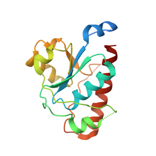
Many Gram-positive and Gram-negative bacteria utilize polysaccharide surface layers called capsules to evade the immune system; consequently, the synthesis and export of the capsule are a potential therapeutic target. In Escherichia coli K-30, the integral membrane tyrosine autokinase Wzc and the cognate phosphatase Wzb have been shown to be key for both synthesis and assembly of capsular polysaccharides. In the Gram-positive bacterium Streptococcus pneumoniae, the CpsCD complex is analogous to Wzc and the phosphatase CpsB is the corresponding cognate phosphatase. The phosphatases are known to dephosphorylate their corresponding autokinases, yet despite their functional equivalence, they share no sequence homology. We present the structure of Wzb in complex with phosphate and high-resolution structures of apo-CpsB and a phosphate-complexed CpsB. We show that both proteins are active toward Wzc and thereby demonstrate that CpsB is not specific for CpsCD. CpsB is a novel enzyme and represents the first solved structure of a tyrosine phosphatase from a Gram-positive bacterium. Wzb and CpsB have completely different structures, suggesting that they must operate by very different mechanisms. Although the mechanism of Wzb can be inferred from previous studies, CpsB appears to have a tyrosine phosphatase mechanism not observed before. We propose a chemical mechanism for CpsB based on site-directed mutagenesis and structural data.
- Centre for Biomolecular Sciences, The University of St. Andrews, Fife, UK.
Organizational Affiliation: