2WGM | pdb_00002wgm

Complete ion-coordination structure in the rotor ring of Na-dependent F-ATP synthase


Experimental Data Snapshot

  • Method: X-RAY DIFFRACTION
  • Resolution: 2.35 Å
  • R-Value Free: 
    0.245 (Depositor), 0.241 (DCC) 
  • R-Value Work: 
    0.220 (Depositor), 0.217 (DCC) 
  • R-Value Observed: 
    0.222 (Depositor) 

Starting Model: experimental
View more details

wwPDB Validation 3D Report Full Report

Validation slider image for 2WGM

This is version 1.2 of the entry. See complete history

Literature

Complete Ion-Coordination Structure in the Rotor Ring of Na(+)-Dependent F-ATP Synthases.

Meier, T.Krah, A.Bond, P.J.Pogoryelov, D.Diederichs, K.Faraldo-Gomez, J.D.

(2009) J Mol Biology 391: 498

  • DOI: https://doi.org/10.1016/j.jmb.2009.05.082
  • Primary Citation Related Structures: 
    2WGM

  • PubMed Abstract: 

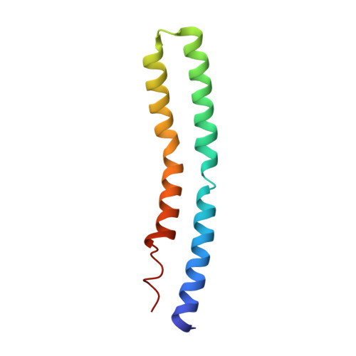
    The membrane-embedded rotors of Na(+)-dependent F-ATP synthases comprise 11 c-subunits that form a ring, with 11 Na(+) binding sites in between adjacent subunits. Following an updated crystallographic analysis of the c-ring from Ilyobacter tartaricus, we report the complete ion-coordination structure of the Na(+) sites. In addition to the four residues previously identified, there exists a fifth ligand, namely, a buried structural water molecule. This water is itself coordinated by Thr67, which, sequence analysis reveals, is the only residue involved in binding that distinguishes Na(+) synthases from H(+)-ATP synthases known to date. Molecular dynamics simulations and free-energy calculations of the c-ring in a lipid membrane lend clear support to the notion that this fifth ligand is a water molecule, and illustrate its influence on the selectivity of the binding sites. Given the evolutionary ascendancy of sodium over proton bioenergetics, this structure uncovers an ancient strategy for selective ion coupling in ATP synthases.


  • Organizational Affiliation
    • Department of Structural Biology, Max Planck Institute of Biophysics, Frankfurt am Main, Germany. thomas.meier@biophys.mpg.de

Macromolecule Content 

  • Total Structure Weight: 394.53 kDa 
  • Atom Count: 28,001 
  • Modeled Residue Count: 3,916 
  • Deposited Residue Count: 3,916 
  • Unique protein chains: 1

Macromolecules

Find similar proteins by:|  3D Structure
Entity ID: 1
MoleculeChains  Sequence LengthOrganismDetailsImage
ATP SYNTHASE SUBUNIT C, SODIUM ION SPECIFIC89Ilyobacter tartaricusMutation(s): 0 
Membrane Entity: Yes 
UniProt
Find proteins for Q8KRV3 (Ilyobacter tartaricus)
Explore Q8KRV3 
Go to UniProtKB:  Q8KRV3
Entity Groups
Sequence Clusters30% Identity50% Identity70% Identity90% Identity95% Identity100% Identity
UniProt GroupQ8KRV3
Sequence Annotations
Expand
Reference Sequence

Small Molecules

Ligands 2 Unique
IDChains Name / Formula / InChI Key2D Diagram3D Interactions
F09

Query on F09


Download:Ideal Coordinates CCD File
BB [auth E]
BC [auth R]
BD [auth i]
BE [auth v]
DB [auth F]
BB [auth E],
BC [auth R],
BD [auth i],
BE [auth v],
DB [auth F],
DC [auth S],
DD [auth j],
FB [auth G],
FC [auth T],
FD [auth k],
HB [auth H],
HC [auth U],
HD [auth l],
JB [auth I],
JC [auth V],
JD [auth m],
LB [auth J],
LC [auth a],
LD [auth n],
NB [auth K],
NC [auth b],
ND [auth o],
PB [auth L],
PC [auth c],
PD [auth p],
RB [auth M],
RC [auth d],
RD [auth q],
TA [auth A],
TB [auth N],
TC [auth e],
TD [auth r],
VA [auth B],
VB [auth O],
VC [auth f],
VD [auth s],
XA [auth C],
XB [auth P],
XC [auth g],
XD [auth t],
ZA [auth D],
ZB [auth Q],
ZC [auth h],
ZD [auth u]
NONAN-1-OL
C9 H20 O
ZWRUINPWMLAQRD-UHFFFAOYSA-N
NA

Query on NA


Download:Ideal Coordinates CCD File
AB [auth E]
AC [auth R]
AD [auth i]
AE [auth v]
CB [auth F]
AB [auth E],
AC [auth R],
AD [auth i],
AE [auth v],
CB [auth F],
CC [auth S],
CD [auth j],
EB [auth G],
EC [auth T],
ED [auth k],
GB [auth H],
GC [auth U],
GD [auth l],
IB [auth I],
IC [auth V],
ID [auth m],
KB [auth J],
KC [auth a],
KD [auth n],
MB [auth K],
MC [auth b],
MD [auth o],
OB [auth L],
OC [auth c],
OD [auth p],
QB [auth M],
QC [auth d],
QD [auth q],
SA [auth A],
SB [auth N],
SC [auth e],
SD [auth r],
UA [auth B],
UB [auth O],
UC [auth f],
UD [auth s],
WA [auth C],
WB [auth P],
WC [auth g],
WD [auth t],
YA [auth D],
YB [auth Q],
YC [auth h],
YD [auth u]
SODIUM ION
Na
FKNQFGJONOIPTF-UHFFFAOYSA-N

Experimental Data & Validation

Experimental Data

  • Method: X-RAY DIFFRACTION
  • Resolution: 2.35 Å
  • R-Value Free:  0.245 (Depositor), 0.241 (DCC) 
  • R-Value Work:  0.220 (Depositor), 0.217 (DCC) 
  • R-Value Observed: 0.222 (Depositor) 
Space Group: P 1 21 1
Unit Cell:
Length ( Å )Angle ( ˚ )
a = 146.69α = 90
b = 139.28β = 118.4
c = 151.91γ = 90
Software Package:
Software NamePurpose
PHENIXrefinement
XDSdata reduction
XSCALEdata scaling
PHASERphasing

Structure Validation

View Full Validation Report


Entry History 

Deposition Data

Revision History  (Full details and data files)

  • Version 1.0: 2009-06-09
    Type: Initial release
  • Version 1.1: 2013-05-29
    Changes: Atomic model, Derived calculations, Non-polymer description, Other, Refinement description, Structure summary, Version format compliance
  • Version 1.2: 2023-12-13
    Changes: Data collection, Database references, Derived calculations, Other, Refinement description