2W5Y

Binary Complex of the Mixed Lineage Leukaemia (MLL1) SET Domain with the cofactor product S-Adenosylhomocysteine.


Experimental Data Snapshot

  • Method: X-RAY DIFFRACTION
  • Resolution: 2.00 Å
  • R-Value Free: 0.264 
  • R-Value Work: 0.187 
  • R-Value Observed: 0.191 

wwPDB Validation   3D Report Full Report


Ligand Structure Quality Assessment 


This is version 1.3 of the entry. See complete history


Literature

Structural Basis for the Requirement of Additional Factors for Mll1 Set Domain Activity and Recognition of Epigenetic Marks.

Southall, S.M.Wong, P.S.Odho, Z.Roe, S.M.Wilson, J.R.

(2009) Mol Cell 33: 181

  • DOI: https://doi.org/10.1016/j.molcel.2008.12.029
  • Primary Citation of Related Structures:  
    2W5Y, 2W5Z

  • PubMed Abstract: 

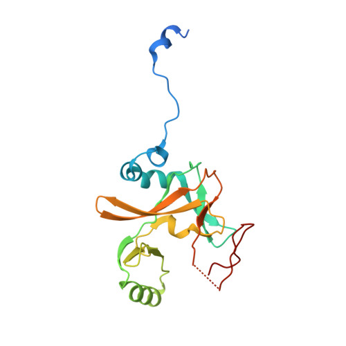
    The mixed-lineage leukemia protein MLL1 is a transcriptional regulator with an essential role in early development and hematopoiesis. The biological function of MLL1 is mediated by the histone H3K4 methyltransferase activity of the carboxyl-terminal SET domain. We have determined the crystal structure of the MLL1 SET domain in complex with cofactor product AdoHcy and a histone H3 peptide. This structure indicates that, in order to form a well-ordered active site, a highly variable but essential component of the SET domain must be repositioned. To test this idea, we compared the effect of the addition of MLL complex members on methyltransferase activity and show that both RbBP5 and Ash2L but not Wdr5 stimulate activity. Additionally, we have determined the effect of posttranslational modifications on histone H3 residues downstream and upstream from the target lysine and provide a structural explanation for why H3T3 phosphorylation and H3K9 acetylation regulate activity.


  • Organizational Affiliation

    Institute of Cancer Research, Chester Beatty Laboratories, Chelsea, London, UK.


Macromolecules
Find similar proteins by:  (by identity cutoff)  |  3D Structure
Entity ID: 1
MoleculeChains Sequence LengthOrganismDetailsImage
HISTONE-LYSINE N-METHYLTRANSFERASE HRX192Homo sapiensMutation(s): 0 
EC: 2.1.1.43
UniProt & NIH Common Fund Data Resources
Find proteins for Q03164 (Homo sapiens)
Explore Q03164 
Go to UniProtKB:  Q03164
PHAROS:  Q03164
GTEx:  ENSG00000118058 
Entity Groups  
Sequence Clusters30% Identity50% Identity70% Identity90% Identity95% Identity100% Identity
UniProt GroupQ03164
Sequence Annotations
Expand
  • Reference Sequence
Small Molecules
Ligands 2 Unique
IDChains Name / Formula / InChI Key2D Diagram3D Interactions
SAH
Query on SAH

Download Ideal Coordinates CCD File 
C [auth A]S-ADENOSYL-L-HOMOCYSTEINE
C14 H20 N6 O5 S
ZJUKTBDSGOFHSH-WFMPWKQPSA-N
ZN
Query on ZN

Download Ideal Coordinates CCD File 
B [auth A]ZINC ION
Zn
PTFCDOFLOPIGGS-UHFFFAOYSA-N
Binding Affinity Annotations 
IDSourceBinding Affinity
SAH BindingDB:  2W5Y IC50: 2300 (nM) from 1 assay(s)
Experimental Data & Validation

Experimental Data

  • Method: X-RAY DIFFRACTION
  • Resolution: 2.00 Å
  • R-Value Free: 0.264 
  • R-Value Work: 0.187 
  • R-Value Observed: 0.191 
  • Space Group: P 21 21 21
Unit Cell:
Length ( Å )Angle ( ˚ )
a = 48.85α = 90
b = 56.21β = 90
c = 78.24γ = 90
Software Package:
Software NamePurpose
PHENIXrefinement
MOSFLMdata reduction
SCALEPACKdata scaling
PHENIXphasing

Structure Validation

View Full Validation Report


Ligand Structure Quality Assessment 


Entry History 

Deposition Data

Revision History  (Full details and data files)

  • Version 1.0: 2009-02-10
    Type: Initial release
  • Version 1.1: 2011-05-08
    Changes: Version format compliance
  • Version 1.2: 2011-07-13
    Changes: Version format compliance
  • Version 1.3: 2023-12-13
    Changes: Data collection, Database references, Derived calculations, Other, Refinement description