2W0Q

E. coli copper amine oxidase in complex with Xenon


Experimental Data Snapshot

  • Method: X-RAY DIFFRACTION
  • Resolution: 2.48 Å
  • R-Value Free: 0.219 
  • R-Value Work: 0.158 
  • R-Value Observed: 0.161 

wwPDB Validation   3D Report Full Report


This is version 1.2 of the entry. See complete history


Literature

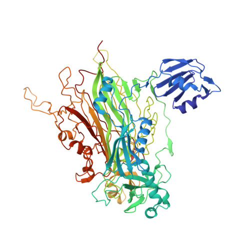
Structure of a Xenon Derivative of Escherichia Coli Copper Amine Oxidase: Confirmation of the Proposed Oxygen-Entry Pathway.

Pirrat, P.Smith, M.A.Pearson, A.R.McPherson, M.J.Phillips, S.E.V.

(2008) Acta Crystallogr Sect F Struct Biol Cryst Commun 64: 1105

  • DOI: https://doi.org/10.1107/S1744309108036373
  • Primary Citation of Related Structures:  
    2W0Q

  • PubMed Abstract: 

    The mechanism of molecular oxygen entry into the buried active site of the copper amine oxidase family has been investigated in several family members using biochemical, structural and in silico methods. These studies have revealed a structurally conserved beta-sandwich which acts as a hydrophobic reservoir from which molecular oxygen can take several species-specific preferred pathways to the active site. Escherichia coli copper amine oxidase (ECAO) possesses an extra N-terminal domain that lies close to one entrance to the beta-sandwich. In order to investigate whether the presence of this domain alters molecular oxygen entry in this enzyme, xenon was used as a molecular oxygen binding-site probe. The resulting 2.5 A resolution X-ray crystal structure reveals xenon bound in similar positions to those observed in xenon-derivative crystal structures of other family members, suggesting that the N-terminal domain does not affect oxygen entry and that the E. coli enzyme takes up oxygen in a similar manner to the rest of the copper amine oxidase family.


  • Organizational Affiliation

    Astbury Centre for Structural Molecular Biology, University of Leeds, Leeds LS2 9JT, England.


Macromolecules
Find similar proteins by:  (by identity cutoff)  |  3D Structure
Entity ID: 1
MoleculeChains Sequence LengthOrganismDetailsImage
COPPER AMINE OXIDASE
A, B
727Escherichia coliMutation(s): 0 
EC: 1.4.3.4
UniProt
Find proteins for P46883 (Escherichia coli (strain K12))
Explore P46883 
Go to UniProtKB:  P46883
Entity Groups  
Sequence Clusters30% Identity50% Identity70% Identity90% Identity95% Identity100% Identity
UniProt GroupP46883
Sequence Annotations
Expand
  • Reference Sequence
Small Molecules
Ligands 3 Unique
IDChains Name / Formula / InChI Key2D Diagram3D Interactions
XE
Query on XE

Download Ideal Coordinates CCD File 
F [auth A]
G [auth A]
H [auth A]
I [auth A]
J [auth A]
F [auth A],
G [auth A],
H [auth A],
I [auth A],
J [auth A],
K [auth A],
O [auth B],
P [auth B],
Q [auth B],
R [auth B],
S [auth B]
XENON
Xe
FHNFHKCVQCLJFQ-UHFFFAOYSA-N
CU
Query on CU

Download Ideal Coordinates CCD File 
C [auth A],
L [auth B]
COPPER (II) ION
Cu
JPVYNHNXODAKFH-UHFFFAOYSA-N
CA
Query on CA

Download Ideal Coordinates CCD File 
D [auth A],
E [auth A],
M [auth B],
N [auth B]
CALCIUM ION
Ca
BHPQYMZQTOCNFJ-UHFFFAOYSA-N
Modified Residues  1 Unique
IDChains TypeFormula2D DiagramParent
TPQ
Query on TPQ
A, B
L-PEPTIDE LINKINGC9 H9 N O5TYR
Experimental Data & Validation

Experimental Data

  • Method: X-RAY DIFFRACTION
  • Resolution: 2.48 Å
  • R-Value Free: 0.219 
  • R-Value Work: 0.158 
  • R-Value Observed: 0.161 
  • Space Group: P 21 21 21
Unit Cell:
Length ( Å )Angle ( ˚ )
a = 134.588α = 90
b = 166.871β = 90
c = 80.33γ = 90
Software Package:
Software NamePurpose
REFMACrefinement
MOSFLMdata reduction
SCALAdata scaling

Structure Validation

View Full Validation Report


Entry History 

Deposition Data

Revision History  (Full details and data files)

  • Version 1.0: 2008-12-16
    Type: Initial release
  • Version 1.1: 2011-05-08
    Changes: Version format compliance
  • Version 1.2: 2011-07-13
    Changes: Version format compliance