2VVI | pdb_00002vvi

IrisFP fluorescent protein in its green form, trans conformation


Experimental Data Snapshot

  • Method: X-RAY DIFFRACTION
  • Resolution: 2.00 Å
  • R-Value Free: 
    0.251 (Depositor) 
  • R-Value Work: 
    0.193 (Depositor) 
  • R-Value Observed: 
    0.196 (Depositor) 

Starting Model: experimental
View more details

wwPDB Validation 3D Report Full Report

Validation slider image for 2VVI

This is version 3.2 of the entry. See complete history

Literature

Structural Characterization of Irisfp, an Optical Highlighter Undergoing Multiple Photo-Induced Transformations.

Adam, V.Lelimousin, M.Boehme, S.Desfonds, G.Nienhaus, K.Field, M.J.Wiedenmann, J.Mcsweeney, S.Nienhaus, G.U.Bourgeois, D.

(2008) Proc Natl Acad Sci U S A 105: 18343

  • DOI: https://doi.org/10.1073/pnas.0805949105
  • Primary Citation Related Structures: 
    2VVH, 2VVI, 2VVJ

  • PubMed Abstract: 

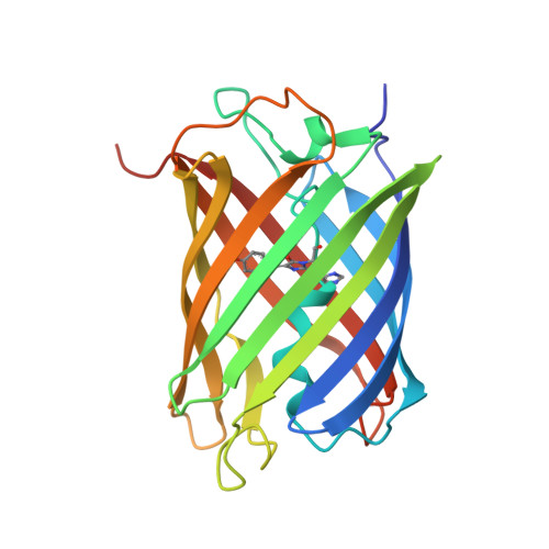
    Photoactivatable fluorescent proteins (FPs) are powerful fluorescent highlighters in live cell imaging and offer perspectives for optical nanoscopy and the development of biophotonic devices. Two types of photoactivation are currently being distinguished, reversible photoswitching between fluorescent and nonfluorescent forms and irreversible photoconversion. Here, we have combined crystallography and (in crystallo) spectroscopy to characterize the Phe-173-Ser mutant of the tetrameric variant of EosFP, named IrisFP, which incorporates both types of phototransformations. In its green fluorescent state, IrisFP displays reversible photoswitching, which involves cis-trans isomerization of the chromophore. Like its parent protein EosFP, IrisFP also photoconverts irreversibly to a red-emitting state under violet light because of an extension of the conjugated pi-electron system of the chromophore, accompanied by a cleavage of the polypeptide backbone. The red form of IrisFP exhibits a second reversible photoswitching process, which may also involve cis-trans isomerization of the chromophore. Therefore, IrisFP displays altogether 3 distinct photoactivation processes. The possibility to engineer and precisely control multiple phototransformations in photoactivatable FPs offers exciting perspectives for the extension of the fluorescent protein toolkit.


  • Organizational Affiliation
    • European Synchrotron Radiation Facility, 6 Rue Jules Horowitz, BP 220, 38043 Grenoble Cedex, France.

Macromolecule Content 

  • Total Structure Weight: 105.82 kDa 
  • Atom Count: 8,289 
  • Modeled Residue Count: 881 
  • Deposited Residue Count: 904 
  • Unique protein chains: 1

Macromolecules

Find similar proteins by:|  3D Structure
Entity ID: 1
MoleculeChains  Sequence LengthOrganismDetailsImage
Green to red photoconvertible GFP-like protein EosFP
A, B, C, D
226Lobophyllia hemprichiiMutation(s): 3 
UniProt
Find proteins for Q5S6Z9 (Lobophyllia hemprichii)
Explore Q5S6Z9 
Go to UniProtKB:  Q5S6Z9
Entity Groups
Sequence Clusters30% Identity50% Identity70% Identity90% Identity95% Identity100% Identity
UniProt GroupQ5S6Z9
Sequence Annotations
Expand
Reference Sequence

Small Molecules

Ligands 2 Unique
IDChains Name / Formula / InChI Key2D Diagram3D Interactions
SO4

Query on SO4


Download:Ideal Coordinates CCD File
E [auth A]
F [auth A]
G [auth A]
I [auth A]
J [auth A]
E [auth A],
F [auth A],
G [auth A],
I [auth A],
J [auth A],
K [auth B],
L [auth B],
M [auth B],
N [auth B],
P [auth C],
Q [auth C],
R [auth C],
T [auth D],
U [auth D],
V [auth D],
W [auth D]
SULFATE ION
O4 S
QAOWNCQODCNURD-UHFFFAOYSA-L
SO3

Query on SO3


Download:Ideal Coordinates CCD File
H [auth A],
O [auth B],
S [auth C],
X [auth D]
SULFITE ION
O3 S
LSNNMFCWUKXFEE-UHFFFAOYSA-L
Modified Residues  1 Unique
IDChains TypeFormula2D DiagramParent
7R0
Query on 7R0
A, B, C, D
L-PEPTIDE LINKINGC17 H17 N5 O4HIS, TYR, GLY

Experimental Data & Validation

Experimental Data

  • Method: X-RAY DIFFRACTION
  • Resolution: 2.00 Å
  • R-Value Free:  0.251 (Depositor) 
  • R-Value Work:  0.193 (Depositor) 
  • R-Value Observed: 0.196 (Depositor) 
Space Group: P 21 21 21
Unit Cell:
Length ( Å )Angle ( ˚ )
a = 81.31α = 90
b = 96.38β = 90
c = 140.61γ = 90
Software Package:
Software NamePurpose
REFMACrefinement
XDSdata reduction
XSCALEdata scaling
PHASERphasing

Structure Validation

View Full Validation Report


Entry History 

Deposition Data

Revision History  (Full details and data files)

  • Version 1.0: 2008-11-11
    Type: Initial release
  • Version 1.1: 2011-05-08
    Changes: Version format compliance
  • Version 1.2: 2011-07-13
    Changes: Version format compliance
  • Version 1.3: 2019-03-06
    Changes: Data collection, Derived calculations, Experimental preparation, Other
  • Version 1.4: 2019-10-23
    Changes: Data collection, Database references, Other
  • Version 2.0: 2021-09-01
    Changes: Atomic model, Data collection, Database references, Derived calculations, Non-polymer description, Polymer sequence, Source and taxonomy, Structure summary
  • Version 3.0: 2023-11-15
    Changes: Atomic model, Data collection, Derived calculations
  • Version 3.1: 2023-12-13
    Changes: Refinement description
  • Version 3.2: 2024-11-13
    Changes: Structure summary