2VS7 | pdb_00002vs7

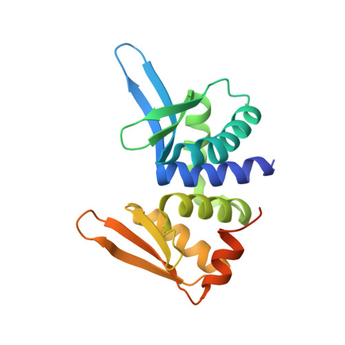
The crystal structure of I-DmoI in complex with DNA and Ca


Experimental Data Snapshot

  • Method: X-RAY DIFFRACTION
  • Resolution: 2.05 Å
  • R-Value Free: 
    0.228 (Depositor), 0.221 (DCC) 
  • R-Value Work: 
    0.187 (Depositor), 0.182 (DCC) 
  • R-Value Observed: 
    0.189 (Depositor) 

wwPDB Validation 3D Report Full Report

Validation slider image for 2VS7

This is version 1.3 of the entry. See complete history. 

Literature

Crystal Structure of I-Dmoi in Complex with its Target DNA Provides New Insights Into Meganuclease Engineering.

Marcaida, M.J., Prieto, J., Redondo, P., Nadra, A.D., Alibes, A., Serrano, L., Grizot, S., Duchateau, P., Paques, F., Blanco, F.J., Montoya, G.

(2008) Proc Natl Acad Sci U S A 105: 16888

  • DOI: https://doi.org/10.1073/pnas.0804795105
  • Primary Citation Related Structures: 
    2VS7, 2VS8

  • PubMed Abstract: 

    Homing endonucleases, also known as meganucleases, are sequence-specific enzymes with large DNA recognition sites. These enzymes can be used to induce efficient homologous gene targeting in cells and plants, opening perspectives for genome engineering with applications in a wide series of fields, ranging from biotechnology to gene therapy. Here, we report the crystal structures at 2.0 and 2.1 A resolution of the I-DmoI meganuclease in complex with its substrate DNA before and after cleavage, providing snapshots of the catalytic process. Our study suggests that I-DmoI requires only 2 cations instead of 3 for DNA cleavage. The structure sheds light onto the basis of DNA binding, indicating key residues responsible for nonpalindromic target DNA recognition. In silico and in vivo analysis of the I-DmoI DNA cleavage specificity suggests that despite the relatively few protein-base contacts, I-DmoI is highly specific when compared with other meganucleases. Our data open the door toward the generation of custom endonucleases for targeted genome engineering using the monomeric I-DmoI scaffold.


  • Organizational Affiliation: 
    • Macromolecular Crystallography and Nuclear Magnetic Resonance Groups, Structural Biology and Biocomputing Programme, Spanish National Cancer Research Centre, c/Melchor Fdez. Almagro 3, 28029 Madrid, Spain.

Macromolecule Content 

  • Total Structure Weight: 116.06 kDa 
  • Atom Count: 8,342 
  • Modeled Residue Count: 697 
  • Deposited Residue Count: 747 
  • Unique protein chains: 1
  • Unique nucleic acid chains: 2

Macromolecules


Find similar proteins by:|  3D Structure
Entity ID: 1
MoleculeChains  Sequence LengthOrganismDetailsImage
HOMING ENDONUCLEASE I-DMOI
A, D, G
199Desulfurococcus mucosusMutation(s): 0 
EC: 3.1
UniProt
Find proteins for P21505 (Desulfurococcus mucosus)
Explore P21505 
Go to UniProtKB:  P21505
Entity Groups
Sequence Clusters30% Identity50% Identity70% Identity90% Identity95% Identity100% Identity
UniProt GroupP21505
Sequence Annotations
Expand
Reference Sequence
Find similar nucleic acids by:  Sequence
Entity ID: 2
MoleculeChains LengthOrganismImage
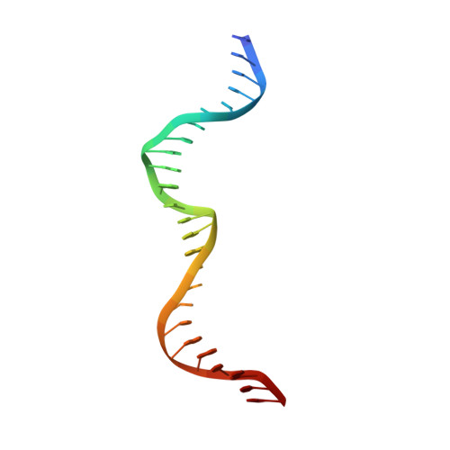
5'-D(*GP*CP*CP*TP*TP*GP*CP*CP*GP*GP *GP*TP*AP*AP*GP*TP*TP*CP*CP*GP*GP*CP*GP*CP*G)-3'
B, E, H
25Desulfurococcus mucosus
Sequence Annotations
Expand
Reference Sequence
Find similar nucleic acids by:  Sequence
Entity ID: 3
MoleculeChains LengthOrganismImage
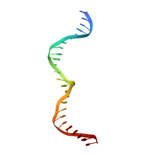
5'-D(*CP*GP*CP*GP*CP*CP*GP*GP*AP*AP *CP*TP*TP*AP*CP*CP*CP*GP*GP*CP*AP*AP*GP*GP*C)-3'
C, F, I
25Desulfurococcus mucosus
Sequence Annotations
Expand
Reference Sequence

Experimental Data & Validation

Experimental Data

  • Method: X-RAY DIFFRACTION
  • Resolution: 2.05 Å
  • R-Value Free:  0.228 (Depositor), 0.221 (DCC) 
  • R-Value Work:  0.187 (Depositor), 0.182 (DCC) 
  • R-Value Observed: 0.189 (Depositor) 
Space Group: P 1 21 1
Unit Cell:
Length ( Å )Angle ( ˚ )
a = 106.988α = 90
b = 70.587β = 119.75
c = 106.848γ = 90
Software Package:
Software NamePurpose
REFMACrefinement
HKL-2000data reduction
SCALEPACKdata scaling
SHARPphasing

Structure Validation

View Full Validation Report


Entry History 

Deposition Data

Revision History  (Full details and data files)

  • Version 1.0: 2008-11-11
    Type: Initial release
  • Version 1.1: 2011-05-08
    Changes: Version format compliance
  • Version 1.2: 2011-07-13
    Changes: Version format compliance
  • Version 1.3: 2024-05-08
    Changes: Data collection, Database references, Derived calculations, Other