2VQ1 | pdb_00002vq1

anti trimeric Lewis X Fab54-5C10-A


Experimental Data Snapshot

  • Method: X-RAY DIFFRACTION
  • Resolution: 2.50 Å
  • R-Value Free: 
    0.260 (Depositor), 0.258 (DCC) 
  • R-Value Work: 
    0.203 (Depositor), 0.202 (DCC) 
  • R-Value Observed: 
    0.206 (Depositor) 

Starting Model: experimental
View more details

wwPDB Validation 3D Report Full Report

Validation slider image for 2VQ1

This is version 1.4 of the entry. See complete history

Literature

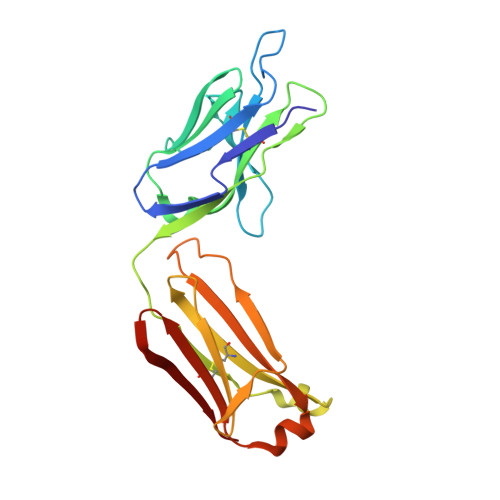
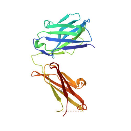
Characterization of a Diagnostic Fab Fragment Binding Trimeric Lewis X.

De Geus, D.C.Van Roon, A.M.M.Thomassen, E.A.J.Hokke, C.H.Deelder, A.M.Abrahams, J.P.

(2009) Proteins 76: 439

  • DOI: https://doi.org/10.1002/prot.22356
  • Primary Citation Related Structures: 
    2VQ1

  • PubMed Abstract: 

    Lewis X trisaccharides normally function as essential cell-cell interaction mediators. However, oligomers of Lewis X trisaccharides expressed by the parasite Schistosoma mansoni seem to be related to its evasion of the immune response of its human host. Here we show that monoclonal antibody 54-5C10-A, which is used to diagnose schistosomiasis in humans, interacts with oligomers of at least three Lewis X trisaccharides, but not with monomeric Lewis X. We describe the sequence and the 2.5 A crystal structure of its Fab fragment and infer a possible mode of binding of the polymeric Lewis X from docking studies. Our studies indicate a radically different mode of binding compared to Fab 291-2G3-A, which is specific for monomeric Lewis X, thus providing a structural explanation of the diagnostic success of 54-5C10-A.


  • Organizational Affiliation
    • Leiden University, The Netherlands. d.de.geus@chem.leidenuniv.nl

Macromolecule Content 

  • Total Structure Weight: 95.7 kDa 
  • Atom Count: 6,868 
  • Modeled Residue Count: 858 
  • Deposited Residue Count: 870 
  • Unique protein chains: 2

Macromolecules

Find similar proteins by:|  3D Structure
Entity ID: 1
MoleculeChains  Sequence LengthOrganismDetailsImage
IGKV1-117 PROTEINA,
C [auth E]
217Mus musculusMutation(s): 0 
UniProt
Find proteins for P01631 (Mus musculus)
Explore P01631 
Go to UniProtKB:  P01631
Find proteins for P01837 (Mus musculus)
Explore P01837 
Go to UniProtKB:  P01837
Entity Groups
Sequence Clusters30% Identity50% Identity70% Identity90% Identity95% Identity100% Identity
UniProt GroupsP01837P01631
Sequence Annotations
Expand
Reference Sequence
Find similar proteins by:|  3D Structure
Entity ID: 2
MoleculeChains  Sequence LengthOrganismDetailsImage
ANTI-HUMAN FC GAMMA RECEPTOR III 3G8 GAMMA HEAVY CHAIN VARIABLE REGIONB,
D [auth F]
218Mus musculusMutation(s): 0 
UniProt
Find proteins for P03987 (Mus musculus)
Explore P03987 
Go to UniProtKB:  P03987
Entity Groups
Sequence Clusters30% Identity50% Identity70% Identity90% Identity95% Identity100% Identity
UniProt GroupP03987
Sequence Annotations
Expand
Reference Sequence

Small Molecules

Ligands 3 Unique
IDChains Name / Formula / InChI Key2D Diagram3D Interactions
GOL

Query on GOL


Download:Ideal Coordinates CCD File
G [auth A],
J [auth B],
P [auth F]
GLYCEROL
C3 H8 O3
PEDCQBHIVMGVHV-UHFFFAOYSA-N
GLY

Query on GLY


Download:Ideal Coordinates CCD File
E [auth A]
F [auth A]
I [auth B]
K [auth E]
L [auth E]
E [auth A],
F [auth A],
I [auth B],
K [auth E],
L [auth E],
M [auth E],
N [auth F],
O [auth F]
GLYCINE
C2 H5 N O2
DHMQDGOQFOQNFH-UHFFFAOYSA-N
AZI

Query on AZI


Download:Ideal Coordinates CCD File
H [auth A]AZIDE ION
N3
IVRMZWNICZWHMI-UHFFFAOYSA-N

Experimental Data & Validation

Experimental Data

  • Method: X-RAY DIFFRACTION
  • Resolution: 2.50 Å
  • R-Value Free:  0.260 (Depositor), 0.258 (DCC) 
  • R-Value Work:  0.203 (Depositor), 0.202 (DCC) 
  • R-Value Observed: 0.206 (Depositor) 
Space Group: P 1 21 1
Unit Cell:
Length ( Å )Angle ( ˚ )
a = 51.384α = 90
b = 161.015β = 103.07
c = 53.48γ = 90
Software Package:
Software NamePurpose
REFMACrefinement
MOSFLMdata reduction
SCALAdata scaling
MOLREPphasing

Structure Validation

View Full Validation Report


Entry History 

Deposition Data

Revision History  (Full details and data files)

  • Version 1.0: 2009-01-27
    Type: Initial release
  • Version 1.1: 2011-06-23
    Changes: Version format compliance
  • Version 1.2: 2011-07-13
    Changes: Version format compliance
  • Version 1.3: 2023-12-13
    Changes: Data collection, Database references, Derived calculations, Other, Refinement description
  • Version 1.4: 2024-11-20
    Changes: Structure summary