Crystal Structure of a Chimera of Human and Plasmodium Falciparum Hypoxanthine Guanine Phosphoribosyltransferases Provides Insights Into Oligomerization.
Gayathri, P., Sujay Subbayya, I.N., Ashok, C.S., Selvi, T.S., Balaram, H., Murthy, M.R.N.(2008) Proteins 73: 1010
- PubMed: 18536021 Search on PubMed
- DOI: https://doi.org/10.1002/prot.22129
- Primary Citation Related Structures:
2VFA - PubMed Abstract:
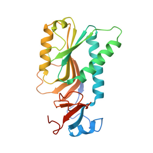
The crystal structure of a chimera of Plasmodium falciparum (Pf) and human hypoxanthine guanine phosphoribosyltransferases (HGPRT), which consists of the core of the protein from the human enzyme and the hood region from the Pf enzyme, has been determined as a complex with the product guanosine monophosphate (GMP). The chimera can utilize hypoxanthine, guanine, and xanthine as substrates, similar to the Pf enzyme. It exists as a monomer-dimer mixture in solution, but shifts to a tetramer on addition of phosphoribosyl pyrophosphate (PRPP). The structural studies reveal that the asymmetric unit of the crystal consists of two monomers of the chimeric HGPRT. Surprisingly, the dimer interface of the chimera is the less extensive AC interface of the parent HGPRTs. An analysis of the crystal structures of the various human HGPRTs provides an explanation for the oligomeric characteristics of the chimera. Pro93 and Tyr197 form part of crucial interactions holding together the AB interface in the unliganded or GMP-bound forms of HGPRT, while Pro93 and His26 interact at the interface after binding of PRPP. Replacement of Tyr197 of human HGPRT by Ile207 in the chimera disrupts the interaction at the AB interface in the absence of PRPP. In the presence of PRPP, the interaction between Pro93 and His26 could restore the AB interface, shifting the chimeric enzyme to a tetrameric state. The structure provides valuable insights into the differences in the AB interface between Pf and human HGPRTs, which may be useful for designing selective inhibitors against the parasite enzyme.
- Molecular Biophysics Unit, Indian Institute of Science, Bangalore 560 012, India.
Organizational Affiliation: