2VCA

Family 89 glycoside hydrolase from Clostridium perfringens in complex with beta-N-acetyl-D-glucosamine


Experimental Data Snapshot

  • Method: X-RAY DIFFRACTION
  • Resolution: 2.05 Å
  • R-Value Free: 0.230 
  • R-Value Work: 0.190 
  • R-Value Observed: 0.192 

wwPDB Validation   3D Report Full Report


Ligand Structure Quality Assessment 


This is version 1.3 of the entry. See complete history


Literature

Structural and Mechanistic Insight Into the Basis of Mucopolysaccharidosis Iiib.

Ficko-Blean, E.Stubbs, K.A.Nemirovsky, O.Vocadlo, D.J.Boraston, A.B.

(2008) Proc Natl Acad Sci U S A 105: 6560

  • DOI: https://doi.org/10.1073/pnas.0711491105
  • Primary Citation of Related Structures:  
    2VC9, 2VCA, 2VCB, 2VCC

  • PubMed Abstract: 

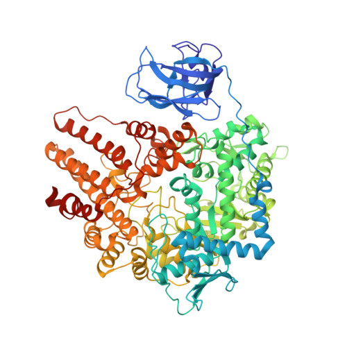
    Mucopolysaccharidosis III (MPS III) has four forms (A-D) that result from buildup of an improperly degraded glycosaminoglycan in lysosomes. MPS IIIB is attributable to the decreased activity of a lysosomal alpha-N-acetylglucosaminidase (NAGLU). Here, we describe the structure, catalytic mechanism, and inhibition of CpGH89 from Clostridium perfringens, a close bacterial homolog of NAGLU. The structure enables the generation of a homology model of NAGLU, an enzyme that has resisted structural studies despite having been studied for >20 years. This model reveals which mutations giving rise to MPS IIIB map to the active site and which map to regions distant from the active site. The identification of potent inhibitors of CpGH89 and the structures of these inhibitors in complex with the enzyme suggest small-molecule candidates for use as chemical chaperones. These studies therefore illuminate the genetic basis of MPS IIIB, provide a clear biochemical rationale for the necessary sequential action of heparan-degrading enzymes, and open the door to the design and optimization of chemical chaperones for treating MPS IIIB.


  • Organizational Affiliation

    Department of Biochemistry and Microbiology, University of Victoria, P.O. Box 3055, Station CSC, Victoria, BC, Canada.


Macromolecules
Find similar proteins by:  (by identity cutoff)  |  3D Structure
Entity ID: 1
MoleculeChains Sequence LengthOrganismDetailsImage
ALPHA-N-ACETYLGLUCOSAMINIDASE891Clostridium perfringensMutation(s): 0 
EC: 3.2.1.50
UniProt
Find proteins for A0A0H2YU91 (Clostridium perfringens (strain ATCC 13124 / DSM 756 / JCM 1290 / NCIMB 6125 / NCTC 8237 / Type A))
Explore A0A0H2YU91 
Go to UniProtKB:  A0A0H2YU91
Entity Groups  
Sequence Clusters30% Identity50% Identity70% Identity90% Identity95% Identity100% Identity
UniProt GroupA0A0H2YU91
Sequence Annotations
Expand
  • Reference Sequence
Experimental Data & Validation

Experimental Data

  • Method: X-RAY DIFFRACTION
  • Resolution: 2.05 Å
  • R-Value Free: 0.230 
  • R-Value Work: 0.190 
  • R-Value Observed: 0.192 
  • Space Group: P 61
Unit Cell:
Length ( Å )Angle ( ˚ )
a = 90.773α = 90
b = 90.773β = 90
c = 252.779γ = 120
Software Package:
Software NamePurpose
REFMACrefinement

Structure Validation

View Full Validation Report


Ligand Structure Quality Assessment 


Entry History 

Deposition Data

Revision History  (Full details and data files)

  • Version 1.0: 2008-03-18
    Type: Initial release
  • Version 1.1: 2011-05-08
    Changes: Version format compliance
  • Version 1.2: 2011-07-13
    Changes: Version format compliance
  • Version 1.3: 2020-07-29
    Type: Remediation
    Reason: Carbohydrate remediation
    Changes: Advisory, Data collection, Derived calculations, Other, Structure summary