2VB7 | pdb_00002vb7

beta-ketoacyl-ACP synthase I (KAS) from E. coli, apo structure after soak in PEG solution


Experimental Data Snapshot

  • Method: X-RAY DIFFRACTION
  • Resolution: 1.60 Å
  • R-Value Free: 
    0.186 (Depositor), 0.205 (DCC) 
  • R-Value Work: 
    0.156 (Depositor) 
  • R-Value Observed: 
    0.158 (Depositor) 

Starting Model: experimental
View more details

wwPDB Validation 3D Report Full Report

Validation slider image for 2VB7

This is version 1.4 of the entry. See complete history

Literature

Structure-Assisted Discovery of an Aminothiazole Derivative as a Lead Molecule for Inhibition of Bacterial Fatty-Acid Synthesis.

Pappenberger, G.Schulz-Gasch, T.Kusznir, E.Mueller, F.Hennig, M.

(2007) Acta Crystallogr D Biol Crystallogr 63: 1208

  • DOI: https://doi.org/10.1107/S0907444907049852
  • Primary Citation Related Structures: 
    2VB7, 2VB8, 2VB9, 2VBA

  • PubMed Abstract: 

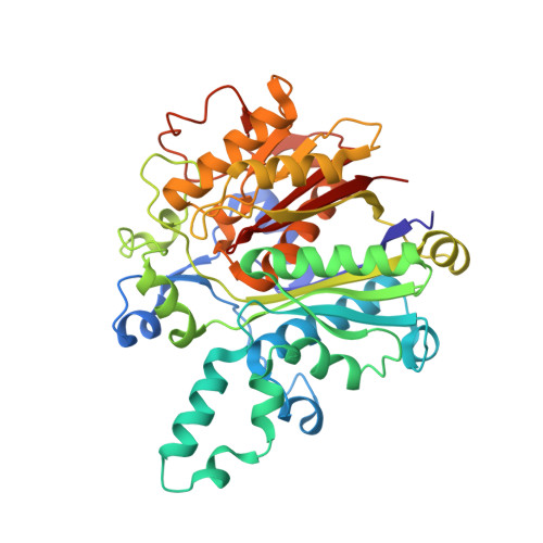
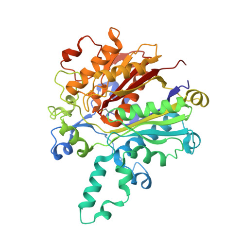
    Fatty-acid synthesis in bacteria is of great interest as a target for the discovery of antibacterial compounds. The addition of a new acetyl moiety to the growing fatty-acid chain, an essential step in this process, is catalyzed by beta-ketoacyl-ACP synthase (KAS). It is inhibited by natural antibiotics such as cerulenin and thiolactomycin; however, these lack the requirements for optimal drug development. Structure-based biophysical screening revealed a novel synthetic small molecule, 2-phenylamino-4-methyl-5-acetylthiazole, that binds to Escherichia coli KAS I with a binding constant of 25 microM as determined by fluorescence titration. A 1.35 A crystal structure of its complex with its target reveals noncovalent interactions with the active-site Cys163 and hydrophobic residues of the fatty-acid binding pocket. The active site is accessible through an open conformation of the Phe392 side chain and no conformational changes are induced at the active site upon ligand binding. This represents a novel binding mode that differs from thiolactomycin or cerulenin interaction. The structural information on the protein-ligand interaction offers strategies for further optimization of this low-molecular-weight compound.


  • Organizational Affiliation
    • F. Hoffmann-La Roche Ltd, Pharma Research Discovery, CH-4070 Basel, Switzerland.

Macromolecule Content 

  • Total Structure Weight: 170.66 kDa 
  • Atom Count: 14,473 
  • Modeled Residue Count: 1,618 
  • Deposited Residue Count: 1,624 
  • Unique protein chains: 2

Macromolecules

Find similar proteins by:|  3D Structure
Entity ID: 1
MoleculeChains  Sequence LengthOrganismDetailsImage
3-OXOACYL-[ACYL-CARRIER-PROTEIN] SYNTHASE 1
A, B, D
406Escherichia coliMutation(s): 0 
EC: 2.3.1.41
UniProt
Find proteins for P0A953 (Escherichia coli (strain K12))
Explore P0A953 
Go to UniProtKB:  P0A953
Entity Groups
Sequence Clusters30% Identity50% Identity70% Identity90% Identity95% Identity100% Identity
UniProt GroupP0A953
Sequence Annotations
Expand
Reference Sequence
Find similar proteins by:|  3D Structure
Entity ID: 2
MoleculeChains  Sequence LengthOrganismDetailsImage
3-OXOACYL-[ACYL-CARRIER-PROTEIN] SYNTHASE 1406Escherichia coliMutation(s): 0 
EC: 2.3.1.41
UniProt
Find proteins for P0A953 (Escherichia coli (strain K12))
Explore P0A953 
Go to UniProtKB:  P0A953
Entity Groups
Sequence Clusters30% Identity50% Identity70% Identity90% Identity95% Identity100% Identity
UniProt GroupP0A953
Sequence Annotations
Expand
Reference Sequence

Small Molecules

Modified Residues  1 Unique
IDChains TypeFormula2D DiagramParent
CSD
Query on CSD
C
L-PEPTIDE LINKINGC3 H7 N O4 SCYS

Experimental Data & Validation

Experimental Data

  • Method: X-RAY DIFFRACTION
  • Resolution: 1.60 Å
  • R-Value Free:  0.186 (Depositor), 0.205 (DCC) 
  • R-Value Work:  0.156 (Depositor) 
  • R-Value Observed: 0.158 (Depositor) 
Space Group: P 21 21 21
Unit Cell:
Length ( Å )Angle ( ˚ )
a = 59.13α = 90
b = 139.46β = 90
c = 212.21γ = 90
Software Package:
Software NamePurpose
REFMACrefinement
XDSdata reduction
XSCALEdata scaling

Structure Validation

View Full Validation Report


Entry History 

Deposition Data

Revision History  (Full details and data files)

  • Version 1.0: 2007-12-25
    Type: Initial release
  • Version 1.1: 2014-11-05
    Changes: Atomic model, Derived calculations, Non-polymer description, Other, Structure summary, Version format compliance
  • Version 1.2: 2019-05-08
    Changes: Data collection, Derived calculations, Experimental preparation, Other
  • Version 1.3: 2023-12-13
    Changes: Data collection, Database references, Other, Refinement description
  • Version 1.4: 2024-11-20
    Changes: Structure summary