2VB1

HEWL at 0.65 angstrom resolution


Experimental Data Snapshot

  • Method: X-RAY DIFFRACTION
  • Resolution: 0.65 Å
  • R-Value Free: 0.095 
  • R-Value Observed: 0.085 

wwPDB Validation   3D Report Full Report


This is version 1.5 of the entry. See complete history


Literature

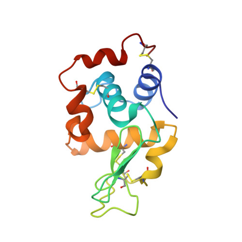
Triclinic Lysozyme at 0.65 A Resolution.

Wang, J.Dauter, M.Alkire, R.Joachimiak, A.Dauter, Z.

(2007) Acta Crystallogr D Biol Crystallogr 63: 1254

  • DOI: https://doi.org/10.1107/S0907444907054224
  • Primary Citation of Related Structures:  
    2VB1

  • PubMed Abstract: 

    The crystal structure of triclinic hen egg-white lysozyme (HEWL) has been refined against diffraction data extending to 0.65 A resolution measured at 100 K using synchrotron radiation. Refinement with anisotropic displacement parameters and with the removal of stereochemical restraints for the well ordered parts of the structure converged with a conventional R factor of 8.39% and an R(free) of 9.52%. The use of full-matrix refinement provided an estimate of the variances in the derived parameters. In addition to the 129-residue protein, a total of 170 water molecules, nine nitrate ions, one acetate ion and three ethylene glycol molecules were located in the electron-density map. Eight sections of the main chain and many side chains were modeled with alternate conformations. The occupancies of the water sites were refined and this step is meaningful when assessed by use of the free R factor. A detailed description and comparison of the structure are made with reference to the previously reported triclinic HEWL structures refined at 0.925 A (at the low temperature of 120 K) and at 0.95 A resolution (at room temperature).


  • Organizational Affiliation

    SAIC-Frederick Inc., Basic Research Program, Argonne National Laboratory, Argonne, IL 60439, USA.


Macromolecules
Find similar proteins by:  (by identity cutoff)  |  3D Structure
Entity ID: 1
MoleculeChains Sequence LengthOrganismDetailsImage
LYSOZYME C129Gallus gallusMutation(s): 0 
EC: 3.2.1.17
UniProt
Find proteins for P00698 (Gallus gallus)
Explore P00698 
Go to UniProtKB:  P00698
Entity Groups  
Sequence Clusters30% Identity50% Identity70% Identity90% Identity95% Identity100% Identity
UniProt GroupP00698
Sequence Annotations
Expand
  • Reference Sequence
Small Molecules
Ligands 3 Unique
IDChains Name / Formula / InChI Key2D Diagram3D Interactions
EDO
Query on EDO

Download Ideal Coordinates CCD File 
C [auth A],
D [auth A],
E [auth A]
1,2-ETHANEDIOL
C2 H6 O2
LYCAIKOWRPUZTN-UHFFFAOYSA-N
NO3
Query on NO3

Download Ideal Coordinates CCD File 
F [auth A]
G [auth A]
H [auth A]
I [auth A]
J [auth A]
F [auth A],
G [auth A],
H [auth A],
I [auth A],
J [auth A],
K [auth A],
L [auth A],
M [auth A],
N [auth A]
NITRATE ION
N O3
NHNBFGGVMKEFGY-UHFFFAOYSA-N
ACT
Query on ACT

Download Ideal Coordinates CCD File 
B [auth A]ACETATE ION
C2 H3 O2
QTBSBXVTEAMEQO-UHFFFAOYSA-M
Experimental Data & Validation

Experimental Data

  • Method: X-RAY DIFFRACTION
  • Resolution: 0.65 Å
  • R-Value Free: 0.095 
  • R-Value Observed: 0.085 
  • Space Group: P 1
Unit Cell:
Length ( Å )Angle ( ˚ )
a = 27.07α = 87.98
b = 31.25β = 108
c = 33.76γ = 112.11
Software Package:
Software NamePurpose
SHELXL-97refinement
HKL-2000data reduction
HKL-2000data scaling
ACORNphasing

Structure Validation

View Full Validation Report


Entry History 

Deposition Data

Revision History  (Full details and data files)

  • Version 1.0: 2007-09-18
    Type: Initial release
  • Version 1.1: 2011-05-08
    Changes: Version format compliance
  • Version 1.2: 2011-07-13
    Changes: Version format compliance
  • Version 1.3: 2019-05-08
    Changes: Data collection, Derived calculations, Experimental preparation, Other
  • Version 1.4: 2019-05-22
    Changes: Data collection, Refinement description
  • Version 1.5: 2023-03-08
    Changes: Database references, Derived calculations, Other