2V9K | pdb_00002v9k

Crystal structure of human PUS10, a novel pseudouridine synthase.


Experimental Data Snapshot

  • Method: X-RAY DIFFRACTION
  • Resolution: 2.00 Å
  • R-Value Free: 
    0.230 (Depositor), 0.227 (DCC) 
  • R-Value Work: 
    0.186 (Depositor), 0.185 (DCC) 
  • R-Value Observed: 
    0.188 (Depositor) 

wwPDB Validation 3D Report Full Report

Validation slider image for 2V9K

This is version 1.2 of the entry. See complete history

Literature

Crystal Structure of Human Pus10, a Novel Pseudouridine Synthase.

Mccleverty, C.J.Hornsby, M.Spraggon, G.Kreusch, A.

(2007) J Mol Biology 373: 1243

  • DOI: https://doi.org/10.1016/j.jmb.2007.08.053
  • Primary Citation Related Structures: 
    2V9K

  • PubMed Abstract: 

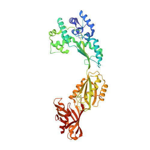
    Pseudouridine (Psi) synthases catalyze the formation of one or more specific Psis in structured RNAs. Five families of Psi synthases have been characterized based on sequence homology. Pus10 has no significant sequence homology to these defined families and therefore represents a new family of Psi synthases. Initial characterization studies show that an archael Pus10 catalyzes the universally conserved Psi55 in tRNA. We present here the crystal structure of human Pus10 at 2.0 A resolution, which is the first structural description from this novel Psi synthase family. Pus10 is a crescent-shaped molecule with two domains, the universally conserved Psi synthase catalytic domain and a THUMP-containing domain, which is unique to the Pus10 family. Superposition of the catalytic domains of Pus10 and other Psi synthases identifies the full set of conserved Psi synthase active site residues indicating that Pus10 likely employs a similar catalytic mechanism to other Psi synthases. The Pus10 active site is located in a deep pocket of a basic cleft adjacent to flexible thumb and forefinger loops, which could provide further stabilization for binding the RNA substrate. Modeling studies demonstrate that the cleft between the catalytic and accessory domain is large enough and electrostatically compatible to accommodate an RNA stem and support the role of the N-terminal domain as an accessory RNA-binding domain.


  • Organizational Affiliation
    • Genomics Institute of the Novartis Research Foundation, 10675 John Jay Hopkins Drive, San Diego, CA 92121, USA.

Macromolecule Content 

  • Total Structure Weight: 61.16 kDa 
  • Atom Count: 4,093 
  • Modeled Residue Count: 467 
  • Deposited Residue Count: 530 
  • Unique protein chains: 1

Macromolecules

Find similar proteins by:|  3D Structure
Entity ID: 1
MoleculeChains  Sequence LengthOrganismDetailsImage
UNCHARACTERIZED PROTEIN FLJ32312530Homo sapiensMutation(s): 0 
EC: 5.4.99.25
UniProt & NIH Common Fund Data Resources
Find proteins for Q3MIT2 (Homo sapiens)
Explore Q3MIT2 
Go to UniProtKB:  Q3MIT2
PHAROS:  Q3MIT2
GTEx:  ENSG00000162927 
Entity Groups
Sequence Clusters30% Identity50% Identity70% Identity90% Identity95% Identity100% Identity
UniProt GroupQ3MIT2
Sequence Annotations
Expand
Reference Sequence

Small Molecules

Ligands 4 Unique
IDChains Name / Formula / InChI Key2D Diagram3D Interactions
EPE

Query on EPE


Download:Ideal Coordinates CCD File
G [auth A],
H [auth A]
4-(2-HYDROXYETHYL)-1-PIPERAZINE ETHANESULFONIC ACID
C8 H18 N2 O4 S
JKMHFZQWWAIEOD-UHFFFAOYSA-N
GOL

Query on GOL


Download:Ideal Coordinates CCD File
I [auth A]GLYCEROL
C3 H8 O3
PEDCQBHIVMGVHV-UHFFFAOYSA-N
ZN

Query on ZN


Download:Ideal Coordinates CCD File
F [auth A]ZINC ION
Zn
PTFCDOFLOPIGGS-UHFFFAOYSA-N
CL

Query on CL


Download:Ideal Coordinates CCD File
B [auth A],
C [auth A],
D [auth A],
E [auth A]
CHLORIDE ION
Cl
VEXZGXHMUGYJMC-UHFFFAOYSA-M

Experimental Data & Validation

Experimental Data

  • Method: X-RAY DIFFRACTION
  • Resolution: 2.00 Å
  • R-Value Free:  0.230 (Depositor), 0.227 (DCC) 
  • R-Value Work:  0.186 (Depositor), 0.185 (DCC) 
  • R-Value Observed: 0.188 (Depositor) 
Space Group: P 32 2 1
Unit Cell:
Length ( Å )Angle ( ˚ )
a = 80.611α = 90
b = 80.611β = 90
c = 153.947γ = 120
Software Package:
Software NamePurpose
REFMACrefinement
DENZOdata reduction
SCALEPACKdata scaling
SOLVEphasing

Structure Validation

View Full Validation Report


Entry History 

Deposition Data

Revision History  (Full details and data files)

  • Version 1.0: 2007-10-09
    Type: Initial release
  • Version 1.1: 2011-07-13
    Changes: Advisory, Refinement description, Version format compliance
  • Version 1.2: 2024-05-08
    Changes: Data collection, Database references, Derived calculations, Other