Structure-Based Drug Design: The Discovery of Novel Nonpeptide Orally Active Inhibitors of Human Renin
Rahuel, J., Rasetti, V., Maibaum, J., Rueger, H., Goschke, R., Cohen, N.C., Stutz, S., Cumin, F., Fuhrer, W., Wood, J.M., Grutter, M.G.(2000) Chem Biol 7: 493
- PubMed: 10903938 Search on PubMed
- DOI: https://doi.org/10.1016/s1074-5521(00)00134-4
- Primary Citation Related Structures:
2V0Z, 2V10, 2V11, 2V12, 2V13, 2V16 - PubMed Abstract:
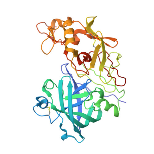
The aspartic proteinase renin plays an important physiological role in the regulation of blood pressure. It catalyses the first step in the conversion of angiotensinogen to the hormone angiotensin II. In the past, potent peptide inhibitors of renin have been developed, but none of these compounds has made it to the end of clinical trials. Our primary aim was to develop novel nonpeptide inhibitors. Based on the available structural information concerning renin-substrate interactions, we synthesized inhibitors in which the peptide portion was replaced by lipophilic moieties that interact with the large hydrophobic S1/S3-binding pocket in renin. Crystal structure analysis of renin-inhibitor complexes combined with computational methods were employed in the medicinal-chemistry optimisation process. Structure analysis revealed that the newly designed inhibitors bind as predicted to the S1/S3 pocket. In addition, however, these compounds interact with a hitherto unrecognised large, distinct, sub-pocket of the enzyme that extends from the S3-binding site towards the hydrophobic core of the enzyme. Binding to this S3(sp) sub-pocket was essential for high binding affinity. This unprecedented binding mode guided the drug-design process in which the mostly hydrophobic interactions within subsite S3(sp) were optimised. Our design approach led to compounds with high in vitro affinity and specificity for renin, favourable bioavailability and excellent oral efficacy in lowering blood pressure in primates. These renin inhibitors are therefore potential therapeutic agents for the treatment of hypertension and related cardiovascular diseases.
- Core Technology Area, Novartis Pharma AG, Metabolic and Cardiovascular Diseases, Basle, Switzerland.
Organizational Affiliation: