2TSB | pdb_00002tsb

AZURIN MUTANT M121A-AZIDE


Experimental Data Snapshot

  • Method: X-RAY DIFFRACTION
  • Resolution: 2.30 Å
  • R-Value Work: 
    0.194 (Depositor) 
  • R-Value Observed: 
    0.194 (Depositor) 

wwPDB Validation 3D Report Full Report

Validation slider image for 2TSB

This is version 1.5 of the entry. See complete history

Literature

Mutant Met121Ala of Pseudomonas aeruginosa azurin and its azide derivative: crystal structures and spectral properties.

Tsai, L.C.Bonander, N.Harata, K.Karlsson, G.Vanngard, T.Langer, V.Sjolin, L.

(1996) Acta Crystallogr D Biol Crystallogr 52: 950-958

  • DOI: https://doi.org/10.1107/S0907444996004982
  • Primary Citation Related Structures: 
    2TSA, 2TSB

  • PubMed Abstract: 

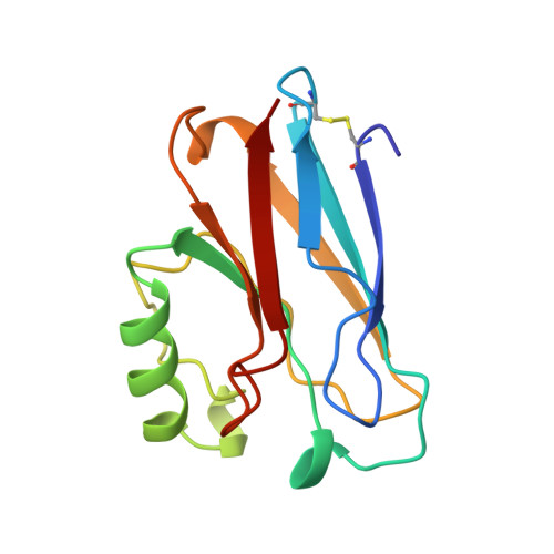
    The crystal structures of the azurin mutant Met121Ala and its azide derivative Met121Ala-azide from Pseudomonas aeruginosa have been determined. The final crystallographic R values are 21.3 and 19.4% for the two structures, respectively. In the Met121Ala mutant, the distance between the copper ion and His117 increases by 0.34 A compared with the wild-type structure. The removal of the methionine in the apical position induces a shortening of the distance from the copper ion to the carbonyl O atom of Gly45 from 2.97 to 2.74 A. In the Met121Ala-azide structure, the azide anion occupies the cavity created by replacing the Met121 side chain with the smaller methyl group of Ala. The azide anion binds with a terminal N atom to the copper ion at a distance of about 2.04 A. In addition, the copper ion has moved out of the trigonal plane by about 0.26 A towards the azide anion. Thus, the copper site in this structure has a distorted tetrahedral arrangement. The spectroscopic characteristics show, in addition, that the copper sites in the two structures are distinctively different. The Met121Ala mutant still maintains the properties of an ordinary type 1 copper site while the Met121Ala-azide derivative has an absorption maximum at about 409 nm and the copper hyperfine coupling has increased to a value intermediate between those of type 2 copper and the wild-type azurin.


  • Organizational Affiliation
    • Department of Inorganic Chemistry, Chalmers University of Technology and Göteborg University, Sweden.

Macromolecule Content 

  • Total Structure Weight: 56.03 kDa 
  • Atom Count: 4,124 
  • Modeled Residue Count: 512 
  • Deposited Residue Count: 512 
  • Unique protein chains: 1

Macromolecules

Find similar proteins by:|  3D Structure
Entity ID: 1
MoleculeChains  Sequence LengthOrganismDetailsImage
AZURIN AZIDE
A, B, C, D
128Pseudomonas aeruginosaMutation(s): 1 
UniProt
Find proteins for P00282 (Pseudomonas aeruginosa (strain ATCC 15692 / DSM 22644 / CIP 104116 / JCM 14847 / LMG 12228 / 1C / PRS 101 / PAO1))
Explore P00282 
Go to UniProtKB:  P00282
Entity Groups
Sequence Clusters30% Identity50% Identity70% Identity90% Identity95% Identity100% Identity
UniProt GroupP00282
Sequence Annotations
Expand
Reference Sequence

Experimental Data & Validation

Experimental Data

  • Method: X-RAY DIFFRACTION
  • Resolution: 2.30 Å
  • R-Value Work:  0.194 (Depositor) 
  • R-Value Observed: 0.194 (Depositor) 
Space Group: P 21 21 21
Unit Cell:
Length ( Å )Angle ( ˚ )
a = 109.7α = 90
b = 98.7β = 90
c = 48.1γ = 90
Software Package:
Software NamePurpose
MADNESdata collection
X-PLORmodel building
X-PLORrefinement
MADNESdata reduction
X-PLORphasing

Structure Validation

View Full Validation Report


Entry History 

Deposition Data

Revision History  (Full details and data files)

  • Version 1.0: 1996-11-08
    Type: Initial release
  • Version 1.1: 2008-03-25
    Changes: Version format compliance
  • Version 1.2: 2011-07-13
    Changes: Version format compliance
  • Version 1.3: 2018-04-18
    Changes: Data collection, Other
  • Version 1.4: 2021-11-03
    Changes: Database references, Derived calculations
  • Version 1.5: 2024-10-16
    Changes: Data collection, Structure summary