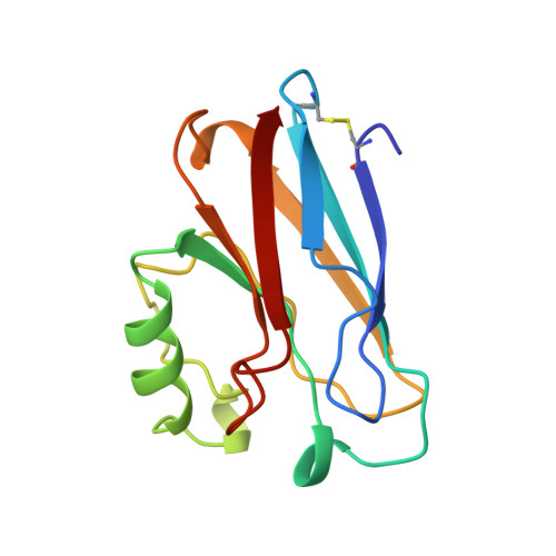
Mutant Met121Ala of Pseudomonas aeruginosa azurin and its azide derivative: crystal structures and spectral properties.
Tsai, L.C., Bonander, N., Harata, K., Karlsson, G., Vanngard, T., Langer, V., Sjolin, L.(1996) Acta Crystallogr D Biol Crystallogr 52: 950-958
- PubMed: 15299604
- DOI: https://doi.org/10.1107/S0907444996004982
- Primary Citation of Related Structures:
2TSA, 2TSB - PubMed Abstract:
The crystal structures of the azurin mutant Met121Ala and its azide derivative Met121Ala-azide from Pseudomonas aeruginosa have been determined. The final crystallographic R values are 21.3 and 19.4% for the two structures, respectively. In the Met121Ala mutant, the distance between the copper ion and His117 increases by 0.34 A compared with the wild-type structure. The removal of the methionine in the apical position induces a shortening of the distance from the copper ion to the carbonyl O atom of Gly45 from 2.97 to 2.74 A. In the Met121Ala-azide structure, the azide anion occupies the cavity created by replacing the Met121 side chain with the smaller methyl group of Ala. The azide anion binds with a terminal N atom to the copper ion at a distance of about 2.04 A. In addition, the copper ion has moved out of the trigonal plane by about 0.26 A towards the azide anion. Thus, the copper site in this structure has a distorted tetrahedral arrangement. The spectroscopic characteristics show, in addition, that the copper sites in the two structures are distinctively different. The Met121Ala mutant still maintains the properties of an ordinary type 1 copper site while the Met121Ala-azide derivative has an absorption maximum at about 409 nm and the copper hyperfine coupling has increased to a value intermediate between those of type 2 copper and the wild-type azurin.
- Department of Inorganic Chemistry, Chalmers University of Technology and Göteborg University, Sweden.
Organizational Affiliation:


