High resolution structure of the N-terminal domain of tissue inhibitor of metalloproteinases-2 and characterization of its interaction site with matrix metalloproteinase-3.
Muskett, F.W., Frenkiel, T.A., Feeney, J., Freedman, R.B., Carr, M.D., Williamson, R.A.(1998) J Biol Chem 273: 21736-21743
- PubMed: 9705310 Search on PubMed
- DOI: https://doi.org/10.1074/jbc.273.34.21736
- Primary Citation Related Structures:
2TMP - PubMed Abstract:
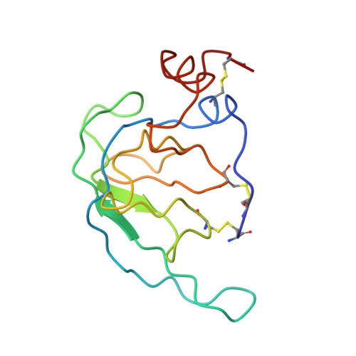
The high resolution structure of the N-terminal domain of tissue inhibitor of metalloproteinases-2 (N-TIMP-2) in solution has been determined using multidimensional heteronuclear NMR spectroscopy, with the structural calculations based on an extensive set of constraints, including 3132 nuclear Overhauser effect-based distance constraints, 56 hydrogen bond constraints, and 220 torsion angle constraints (an average of 26.9 constraints/residue). The core of the protein consists of a five-stranded beta-barrel that is homologous to the beta-barrel found in the oligosaccharide/oligonucleotide binding protein fold. The binding site for the catalytic domain of matrix metalloproteinases-3 (N-MMP-3) on N-TIMP-2 has been mapped by determining the changes in chemical shifts on complex formation for signals from the protein backbone (15N, 13C, and 1H). This approach identified a discrete N-MMP-3 binding site on N-TIMP-2 composed of the N terminus of the protein and the loops between beta-strands AB, CD, and EF. The beta-hairpin formed from strands A and B in N-TIMP-2 is significantly longer than the equivalent structure in TIMP-1, allowing it to make more extensive binding interactions with the MMP catalytic domain. A detailed comparison of the N-TIMP-2 structure with that of TIMP-1 bound to N-MMP-3 (Gomis-Ruth, F.-X., Maskos, K., Betz, M., Bergner, A., Huber, R., Suzuki, K., Yoshida, N., Nagase, H. , Brew, K., Bourne, G. P., Bartunik, H. & Bode, W. (1997) Nature 389, 77-80) revealed that the core beta-barrels are very similar in topology but that the loop connecting beta-strands CD (P67-C72) would need to undergo a large conformational change for TIMP-2 to bind in a similar manner to TIMP-1.
- Department of Biosciences, University of Kent, Canterbury, Kent, CT2 7NJ, United Kingdom.
Organizational Affiliation: