2TMD | pdb_00002tmd

CORRELATION OF X-RAY DEDUCED AND EXPERIMENTAL AMINO ACID SEQUENCES OF TRIMETHYLAMINE DEHYDROGENASE


Experimental Data Snapshot

  • Method: X-RAY DIFFRACTION
  • Resolution: 2.40 Å
  • R-Value Work: 
    0.154 (Depositor) 
  • R-Value Observed: 
    0.154 (Depositor) 

wwPDB Validation 3D Report Full Report

Validation slider image for 2TMD

This is version 1.3 of the entry. See complete history

Literature

Correlation of x-ray deduced and experimental amino acid sequences of trimethylamine dehydrogenase.

Barber, M.J.Neame, P.J.Lim, L.W.White, S.Matthews, F.S.

(1992) J Biological Chem 267: 6611-6619

  • Primary Citation Related Structures: 
    2TMD

  • PubMed Abstract: 

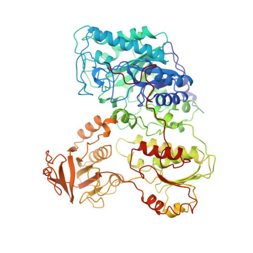
    The amino acid sequence of the iron-sulfur-flavoprotein, trimethylamine dehydrogenase, isolated from the bacterium W3A1 has been deduced from the x-ray diffraction pattern obtained at 2.4-A resolution. This sequence has been compared to portions of the primary sequence derived by gas-phase sequencing of isolated peptides obtained from cyanogen bromide and endoprotease Arg-C and Asp-N digestions of the purified enzyme. A consensus sequence has resulted and is comprised of 729 amino acids with Ala at both NH2- and COOH-terminal positions. The consensus sequence contains 13 cysteine residues. Approximately 80% of the sequence has been confirmed by direct sequencing with approximately 81% agreement with the x-ray deduced sequence. The calculated subunit molecular mass of the apoenzyme is 78,899 Da, in good agreement with published values of approximately 83,000. The anomalous scattering map from the native protein has also been shown to provide accurate information about the positions of most of the weak anomalous scattering centers such as sulfur or phosphorus atoms and to complement x-ray or chemical sequencing methods.


  • Organizational Affiliation
    • Department of Biochemistry and Molecular Biology, University of South Florida, College of Medicine, Tampa 33612.

Macromolecule Content 

  • Total Structure Weight: 165.68 kDa 
  • Atom Count: 12,195 
  • Modeled Residue Count: 1,458 
  • Deposited Residue Count: 1,458 
  • Unique protein chains: 1

Macromolecules

Find similar proteins by:|  3D Structure
Entity ID: 1
MoleculeChains  Sequence LengthOrganismDetailsImage
TRIMETHYLAMINE DEHYDROGENASE
A, B
729Methylophilus methylotrophus W3A1Mutation(s): 0 
EC: 1.5.99.7 (PDB Primary Data), 1.5.8.2 (UniProt)
UniProt
Find proteins for P16099 (Methylophilus methylotrophus)
Explore P16099 
Go to UniProtKB:  P16099
Entity Groups
Sequence Clusters30% Identity50% Identity70% Identity90% Identity95% Identity100% Identity
UniProt GroupP16099
Sequence Annotations
Expand
Reference Sequence

Experimental Data & Validation

Experimental Data

  • Method: X-RAY DIFFRACTION
  • Resolution: 2.40 Å
  • R-Value Work:  0.154 (Depositor) 
  • R-Value Observed: 0.154 (Depositor) 
Space Group: P 1 21 1
Unit Cell:
Length ( Å )Angle ( ˚ )
a = 147.75α = 90
b = 71.95β = 97.69
c = 83.82γ = 90
Software Package:
Software NamePurpose
X-PLORmodel building
X-PLORrefinement
X-PLORphasing

Structure Validation

View Full Validation Report


Entry History 

Deposition Data

Revision History  (Full details and data files)

  • Version 1.0: 1994-01-31
    Type: Initial release
  • Version 1.1: 2008-03-25
    Changes: Version format compliance
  • Version 1.2: 2011-07-13
    Changes: Source and taxonomy, Version format compliance
  • Version 1.3: 2024-02-21
    Changes: Data collection, Database references, Derived calculations, Other