2ROL

Structural Basis of PxxDY motif recognition in SH3 binding


Experimental Data Snapshot

  • Method: SOLUTION NMR
  • Conformers Calculated: 100 
  • Conformers Submitted: 20 
  • Selection Criteria: structures with the least restraint violations 

wwPDB Validation   3D Report Full Report


This is version 1.3 of the entry. See complete history


Literature

Structural basis of PxxDY motif recognition in SH3 binding

Aitio, O.Hellman, M.Kesti, T.Kleino, I.Samuilova, O.Paakkonen, K.Tossavainen, H.Saksela, K.Permi, P.

(2008) J Mol Biol 382: 167-178

  • DOI: https://doi.org/10.1016/j.jmb.2008.07.008
  • Primary Citation of Related Structures:  
    2K2M, 2ROL

  • PubMed Abstract: 

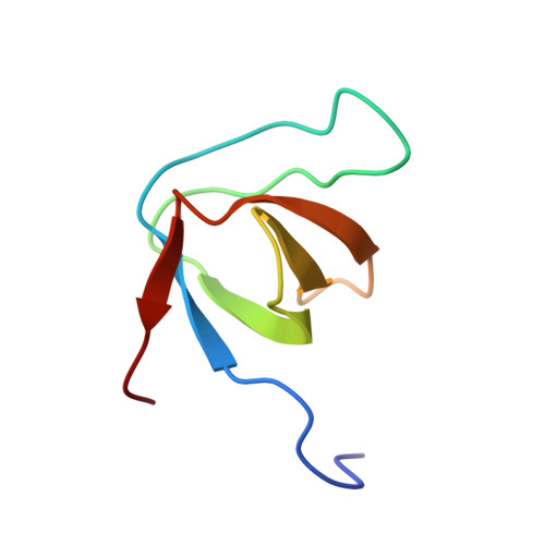
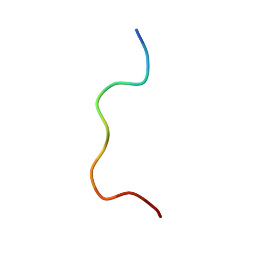
    We have determined the solution structure of epidermal growth factor receptor pathway substrate 8 (Eps8) L1 Src homology 3 (SH3) domain in complex with the PPVPNPDYEPIR peptide from the CD3epsilon cytoplasmic tail. Our structure reveals the distinct structural features that account for the unusual specificity of the Eps8 family SH3 domains for ligands containing a PxxDY motif instead of canonical PxxP ligands. The CD3epsilon peptide binds Eps8L1 SH3 in a class II orientation, but neither adopts a polyproline II helical conformation nor engages the first proline-binding pocket of the SH3 ligand binding interface. Ile531 of Eps8L1 SH3, instead of Tyr or Phe residues typically found in this position in SH3 domains, renders this hydrophobic pocket smaller and nonoptimal for binding to conventional PxxP peptides. A positively charged arginine at position 512 in the n-Src loop of Eps8L1 SH3 plays a key role in PxxDY motif recognition by forming a salt bridge to D7 of the CD3epsilon peptide. In addition, our structural model suggests a hydrogen bond between the hydroxyl group of the aromatic ring of Y8 and the carboxyl group of E496, thus explaining the critical role of the PxxDY motif tyrosine residue in binding to Eps8 family SH3. These finding have direct implications also for understanding the atypical binding specificity of the amino-terminal SH3 of the Nck family proteins.


  • Organizational Affiliation

    Program in Structural Biology and Biophysics, Institute of Biotechnology/NMR Laboratory, University of Helsinki, FI-00014 Helsinki, Finland.


Macromolecules
Find similar proteins by:  (by identity cutoff)  |  3D Structure
Entity ID: 1
MoleculeChains Sequence LengthOrganismDetailsImage
Epidermal growth factor receptor kinase substrate 8-like protein 164Homo sapiensMutation(s): 0 
Gene Names: EPS8L1
UniProt & NIH Common Fund Data Resources
Find proteins for Q8TE68 (Homo sapiens)
Explore Q8TE68 
Go to UniProtKB:  Q8TE68
PHAROS:  Q8TE68
GTEx:  ENSG00000131037 
Entity Groups  
Sequence Clusters30% Identity50% Identity70% Identity90% Identity95% Identity100% Identity
UniProt GroupQ8TE68
Sequence Annotations
Expand
  • Reference Sequence

Find similar proteins by:  Sequence   |   3D Structure  

Entity ID: 2
MoleculeChains Sequence LengthOrganismDetailsImage
12-meric peptide from T-cell surface glycoprotein CD3 epsilon chain12N/AMutation(s): 0 
UniProt & NIH Common Fund Data Resources
Find proteins for P07766 (Homo sapiens)
Explore P07766 
Go to UniProtKB:  P07766
PHAROS:  P07766
GTEx:  ENSG00000198851 
Entity Groups  
Sequence Clusters30% Identity50% Identity70% Identity90% Identity95% Identity100% Identity
UniProt GroupP07766
Sequence Annotations
Expand
  • Reference Sequence
Experimental Data & Validation

Experimental Data

  • Method: SOLUTION NMR
  • Conformers Calculated: 100 
  • Conformers Submitted: 20 
  • Selection Criteria: structures with the least restraint violations 

Structure Validation

View Full Validation Report


Entry History 

Deposition Data

Revision History  (Full details and data files)

  • Version 1.0: 2009-03-03
    Type: Initial release
  • Version 1.1: 2011-07-13
    Changes: Version format compliance
  • Version 1.2: 2020-02-26
    Changes: Data collection, Database references, Derived calculations, Other
  • Version 1.3: 2024-05-01
    Changes: Data collection, Database references